Skip to content

List 99+ Pictures what does an mri of the foot show Updated

List showcases captivating images of what does an mri of the foot show gathered and meticulously curated by the website finwise.edu.vn. Furthermore, you can find more related images in the details below.

what does an mri of the foot show

My Image 1
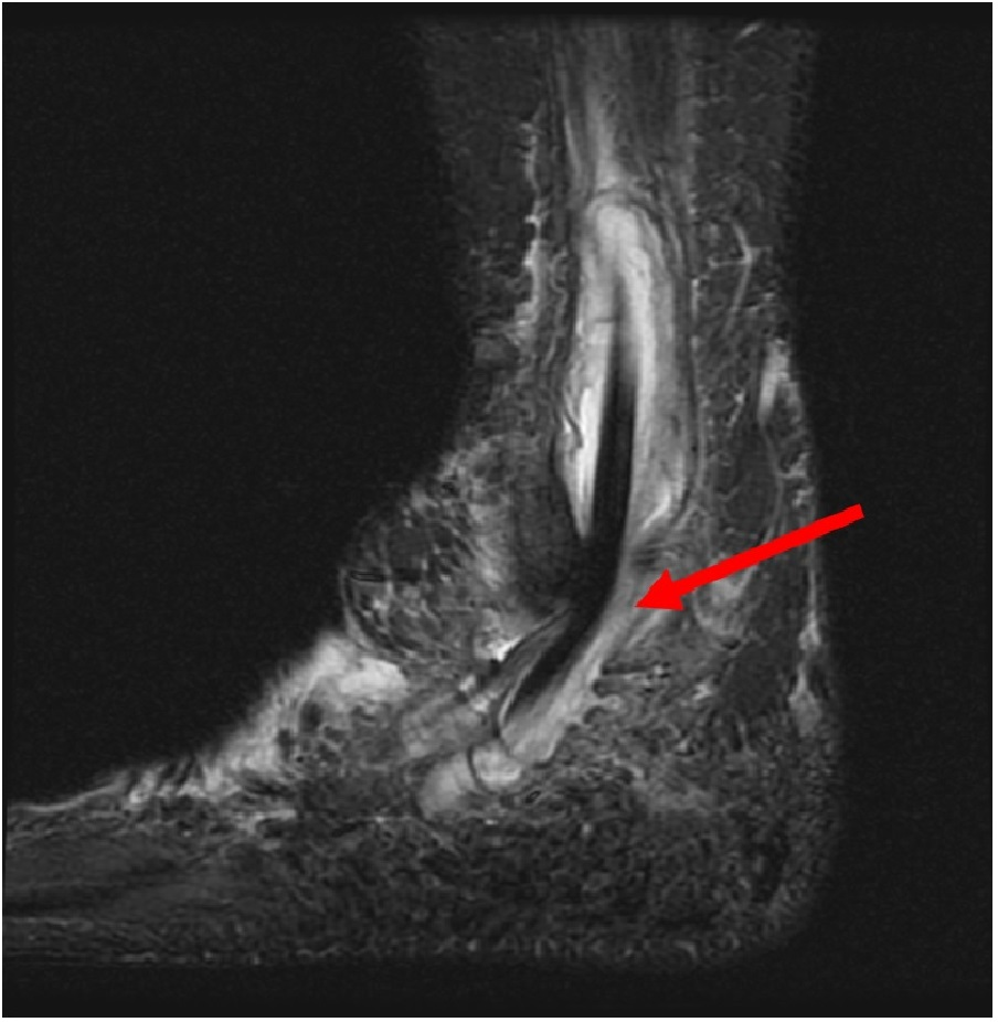
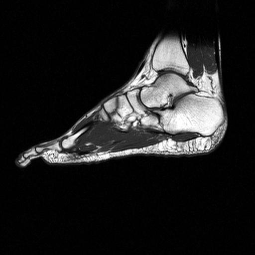
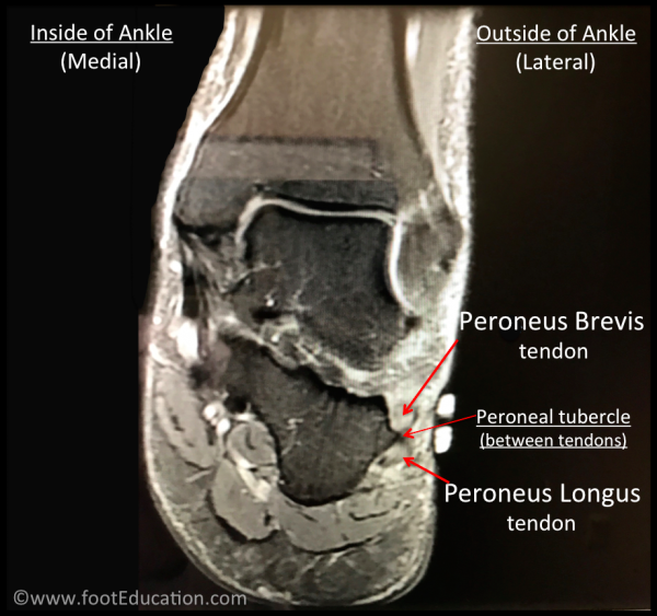
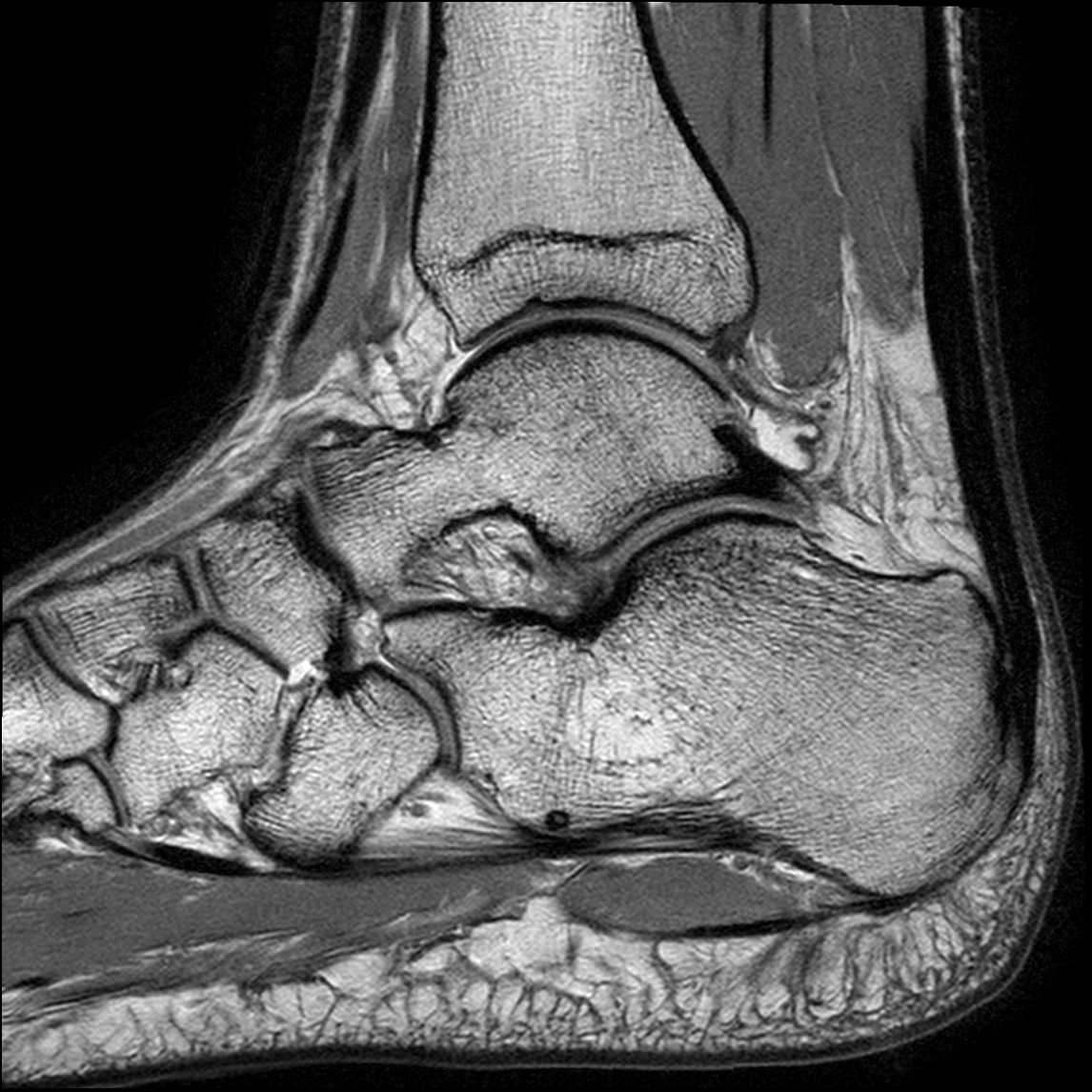
MRI image of ankle and foot showing posterosuperior bony spurring of …
My Image 2
Ankle MRI | Elite MRI of Michigan
My Image 3
G : ‘ mri scan foot’ | Mri, Mri scan, Abstract artwork
My Image 4
Magnetic Resonance Imaging MRI Scan of Foot/Ankle Stock Image – Image …
My Image 5
MRI of the whole foot, sagittal (a) T2W FS, (b &c) T1W FSE pre-and (d …
My Image 6
Cureus | Extrapulmonary Sarcoidosis: A Diagnostic Challenge
My Image 7
Pin on MRI
My Image 8
MRI of Sports Injuries – Musculoskeletal MRI
My Image 9
MRI scan right foot, T2 weighted, 5 days after admission. Marked …
My Image 10
Diffuse Soft Tissue Edema
My Image 11
Cureus | Idiopathic Peroneal Tenosynovitis Caused by a Previously …
My Image 12
Same foot as in Figure 3. Follow-up MRI sagittal STIRsequence shows …
My Image 13
STOCK IMAGE, mri of the ankle sagittal section showing a normal …
My Image 14
MRI; Ankle Synovitis and FHL Tenosynovitis | Download Scientific Diagram
My Image 15
Left foot magnetic resonance imaging. (A) A left foot MRI scan revealed …
My Image 16
Plantar fasciitis on MRI. A sagittal STIR image shows fusiform swelling …
My Image 17
MRI showing nodular exophytic growth right heel with no musculoskeletal …
My Image 18
Normal foot MRI | Image | Radiopaedia.org
My Image 19
and 2. MRI of the foot. Multi-lobe lesion involving only the first …
My Image 20
Rheumatoid nodule of the foot – MRI features | Eurorad
My Image 21
MRI of the left foot. Gadolinium contrast-enhanced axial T1weighted MRI …
My Image 22
MRI foot – radRounds Radiology Network
My Image 23
MRI of the foot (Sagital T2WI) shows a well defined fusiform shape soft …
My Image 24
MRI of the patient’s right foot showing the posterior tibial nerve and …
My Image 25
12 Coronal MRI images of the foot. A abductor hallucis, B flexor …
My Image 26
11 Axial MRI images of the foot. (a) T1-weighted image; (b) T2-weighted …
My Image 27
Neuroma foot mri
My Image 28
(a-d) MRI examination of the foot axial T1 WI post contrast (a), axial …
My Image 29
The Radiology Assistant : Diabetic foot – MRI examination
My Image 30
An Atypical Soft Tissue Mass on the Dorsum of the Foot: A Case Report
My Image 31
Routine MRI findings of the asymptomatic foot in diabetic patients with …
My Image 32
Normal Magnetic Resonance Imaging Anatomy of the Ankle & Foot …
My Image 33
foot mri t1 axial image
My Image 34
MRI of the fore-foot; short-axis (a) T2 FS, sagittal (b) non-contrast …
My Image 35
Read on to find out more about my review areas on a foot X-Ray⁣ ⁣ ⁣ 👨🏽 …
My Image 36
Contract Service MRI – US Occ Docs
My Image 37
T1 weighted axial magnetic resonance imaging (MRI) of the feet showing …
My Image 38
Normal Magnetic Resonance Imaging Anatomy of the Ankle & Foot …
My Image 39
Feet MRI
My Image 40
The Pictures That Show What Covid Is Really Doing To Joints | HuffPost …
My Image 41
Bone SPECT/CT imaging of the painful foot/ankle. Illustration of bone …
My Image 42
MRI of the left foot, shows low signal intensity lesion on the MH. The …
My Image 43
Normal foot MRI | Image | Radiopaedia.org
My Image 44
Foot Muscles Mri / Baxter S Nerve First Branch Of The Lateral Plantar …
My Image 45
Cureus | Idiopathic Peroneal Tenosynovitis Caused by a Previously …
My Image 46
MRI of the right feet: sagittal T1-weighted image demonstrated a …
My Image 47
Foot Muscles Mri – Mri Ankle Anatomy Ankle Anatomy Foot Anatomy Anatomy …
My Image 48
foot mri t1 axial image
My Image 49
Pin on X-rays
My Image 50
hemangioma | The Foot and Ankle Online Journal
My Image 51
tumor of the foot | The Foot and Ankle Online Journal
My Image 52
Heterogeneous signal in bone marrow on MRI of children’s feet: a normal …
My Image 53
Foot Muscles Mri / Mri Scan For Ankle Injury Melbourne Melbourne …
My Image 54
Madura Foot: MRI – Sumer’s Radiology Blog
My Image 55
Foot Muscles Mri Anatomy – Foot anatomy mri coronal Images – The foot …
My Image 56
Foot Muscles Mri Anatomy : anatomy of hip joint | free MRI coronal …
My Image 57
MRI of the right foot -shows diffuse soft tissue swelling with mild …
My Image 58
MRI of the right feet: axial T1-weighted image showing nodular …
My Image 59
Non-contrast fat-saturated T2-weighted coronal MRI of the foot showing …
My Image 60
Best Human Foot X Ray X Ray Image Mri Scan Stock Photos, Pictures …
My Image 61
Ankle/Foot MRI
My Image 62
MRI ankle – Google Search | Ankle anatomy, Mri, Radiology imaging
My Image 63
The Radiology Assistant : Diabetic foot – MRI examination
My Image 64
Rheumatoid nodule of the foot – MRI features | Eurorad
My Image 65
Mobile MRI of the feet. A: male, 39-years-old, stage 53, 3,669 km …
My Image 66
MRI of Foot and Ankle – normal anterior talofibular ligament – MRI at …
My Image 67
Feet MRI
My Image 68
Plantar Foot Muscles Mri / Foot Pain Causes Treatment And When To See A …
My Image 69
foot mri t1 axial image
My Image 70
Foot Muscles Mri / Ultrasound – Feet and ankles ankle muscle anatomy of …
My Image 71
MRI Protocols: ANKLE MRI IMAGING PLANES
My Image 72
Peroneal Tendonitis – FootEducation
My Image 73
Cubo-navicular coalition – a rare cause of chronic foot pain | Eurorad
My Image 74
Foot Muscles Mri : Accessory foot muscle-MRI – Sumer’s Radiology Blog
My Image 75
Ankle and Foot | Radiology Key
My Image 76
foot mri stir sagittal image
My Image 77
mri-achilles-tendon – MRI at Melbourne Radiology Clinic
My Image 78
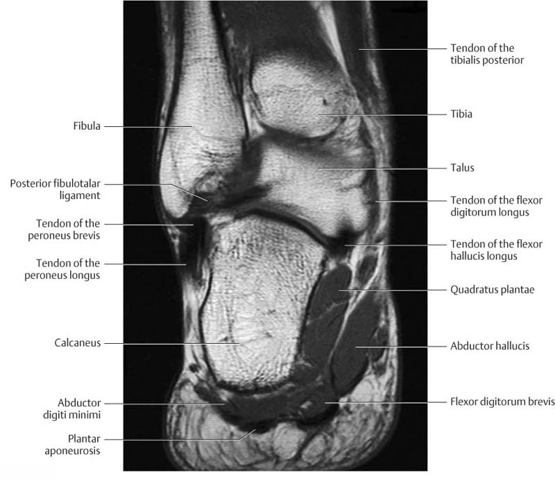
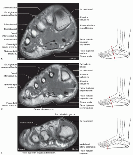
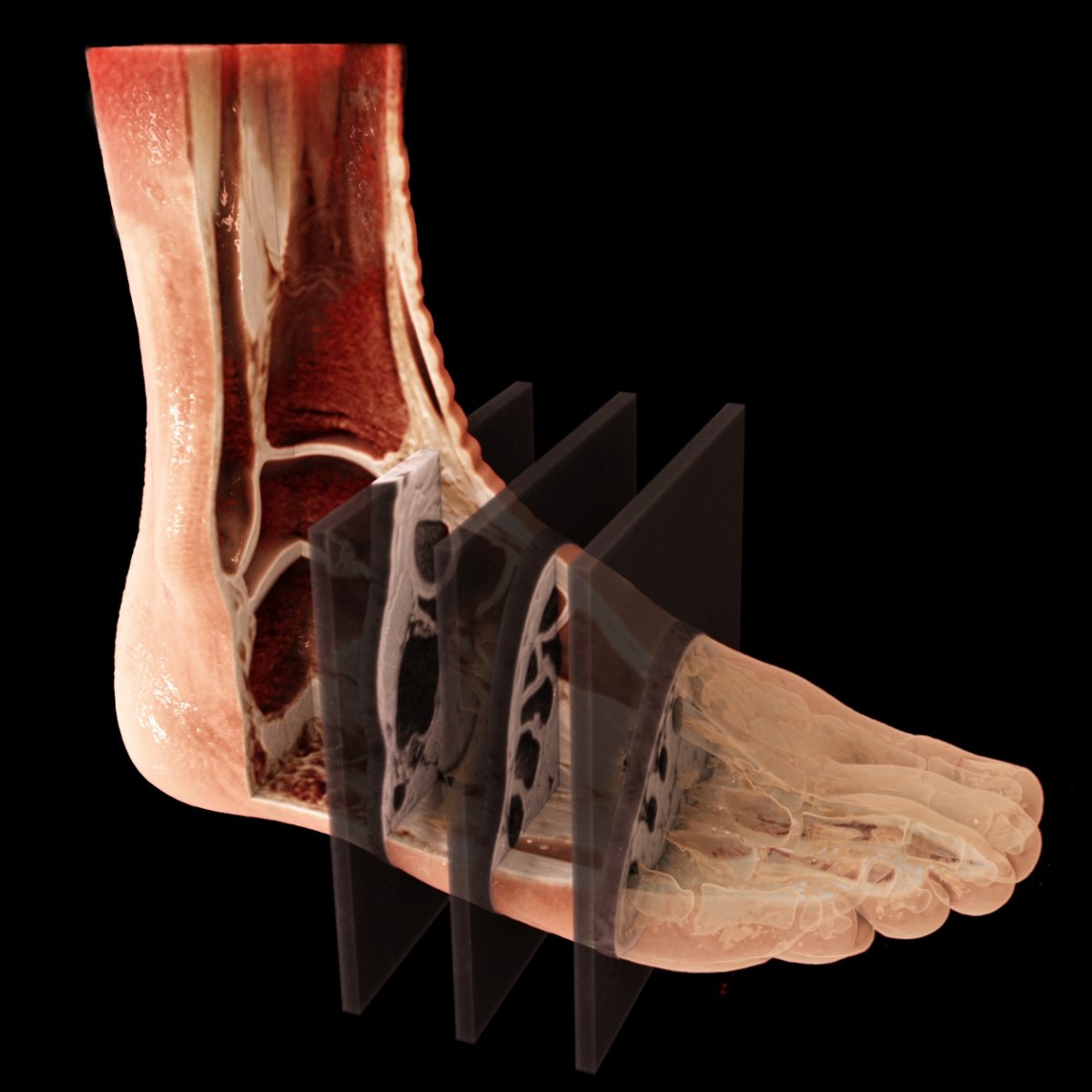
3 D Color MRI Foot Anatomy | Medical websites, Medical, Mri
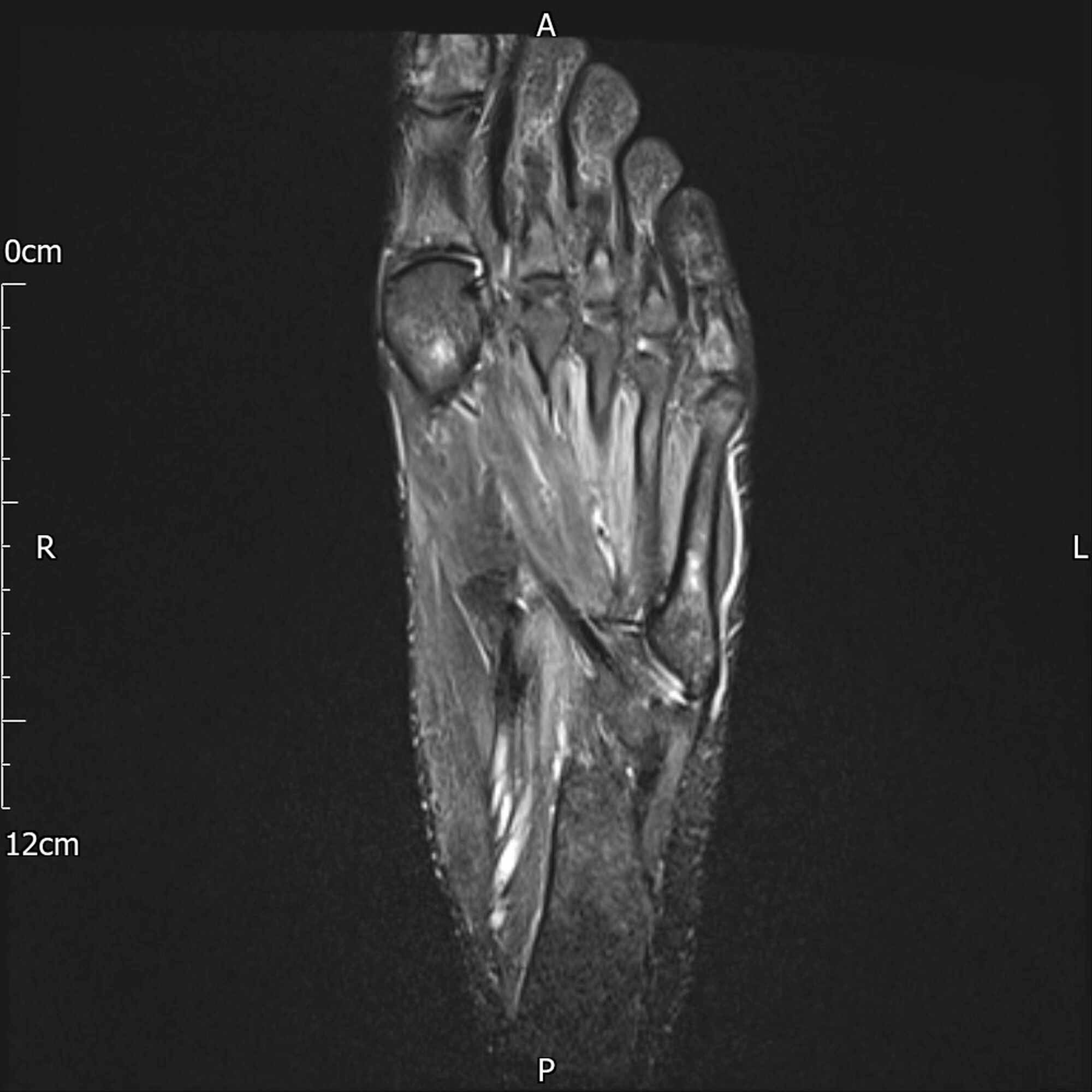
My Image 79
Feet MRI
My Image 80
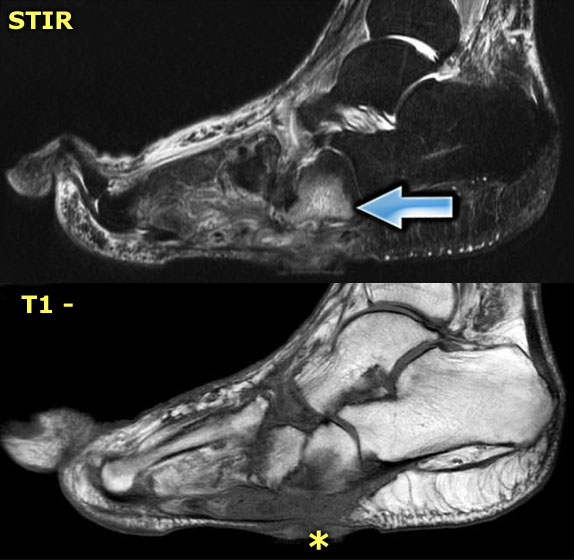
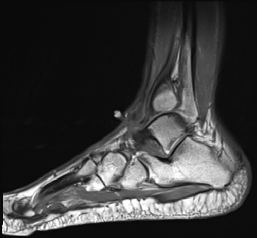
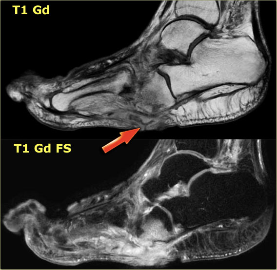
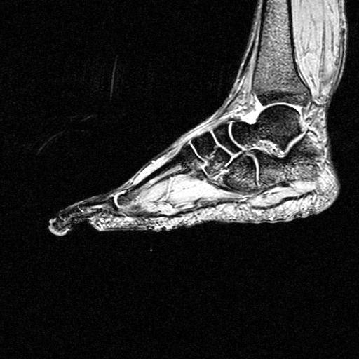
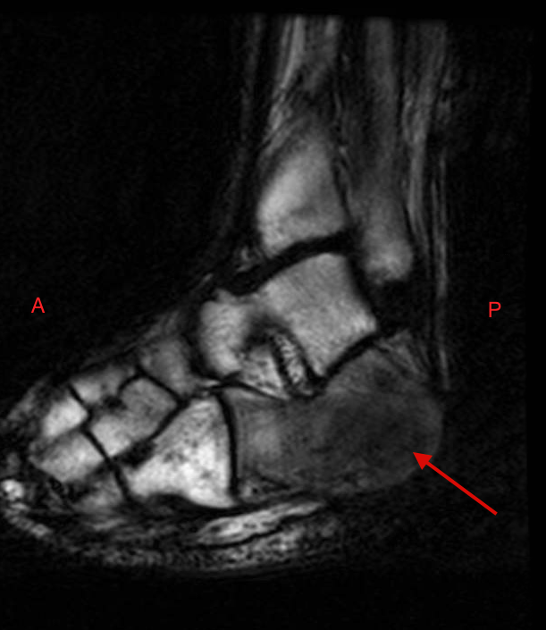
(a) T1-weighted sagittal MRI of the right foot. Note the altered marrow …
My Image 81
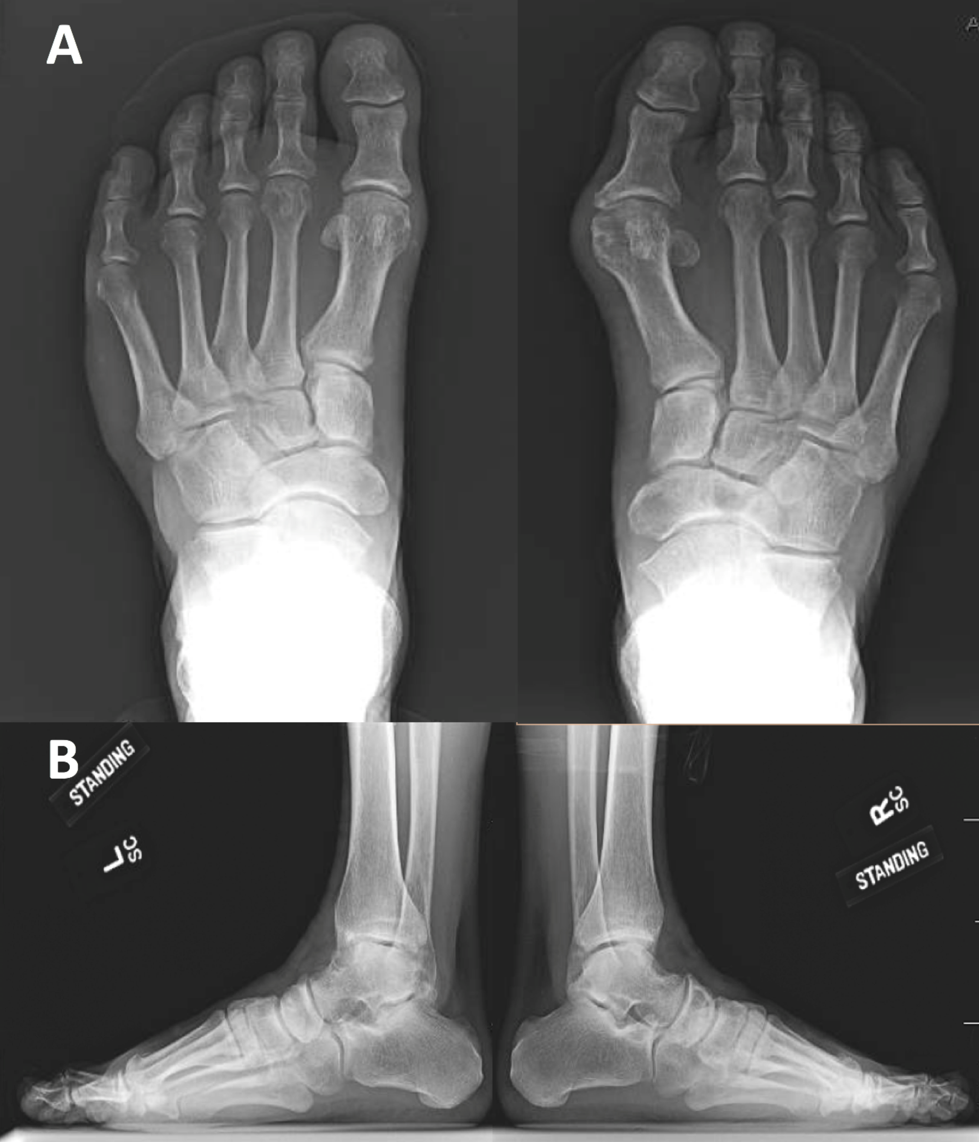
Foot Xray Photos and Premium High Res Pictures – Getty Images
My Image 82
CT, X-ray and MRI of left ankle and foot after 24 months of follow. No …
My Image 83
Giant Intraosseous Synovial Cyst with Intraarticular Communication with …
My Image 84
Benign schwannoma of the medial dorsal cutaneous nerve of the foot: A …
My Image 85
AI helpers simplify clinical MRI scans • healthcare-in-europe.com
My Image 86
MRIs make you feel claustrophobic? An extremity MRI won’t | Catching …
My Image 87
Madura Foot: MRI – Sumer’s Radiology Blog
My Image 88
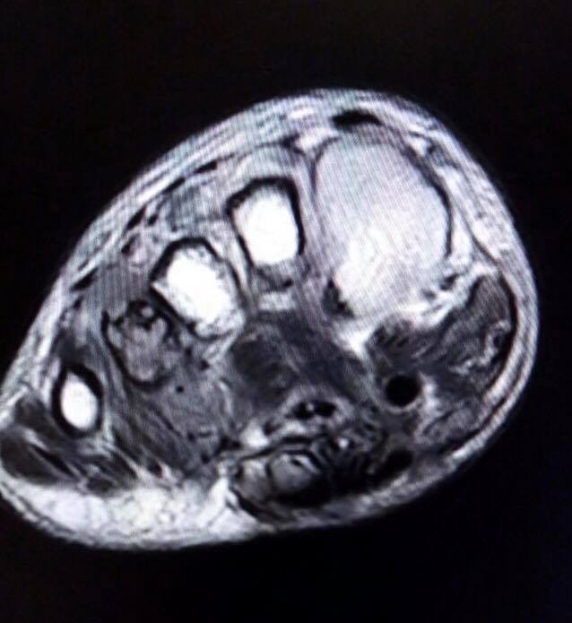
Cureus | A Case of a Second Intermetatarsal Space Gouty Tophus with a …
My Image 89
Image | Radiopaedia.org
My Image 90
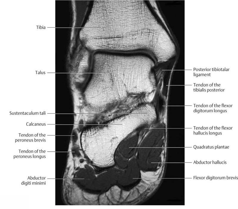
Plantar Foot Muscles Mri – Mri imaging will determine the exact …
My Image 91
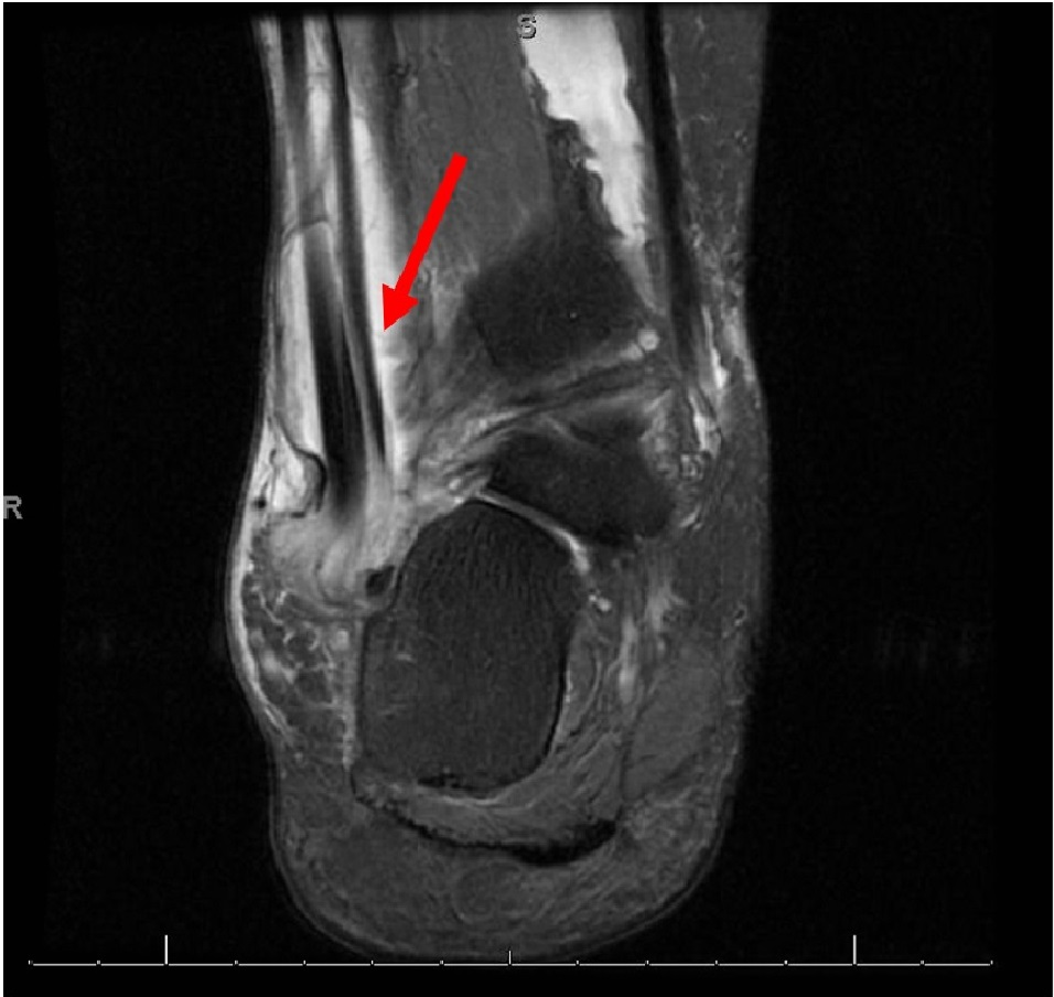
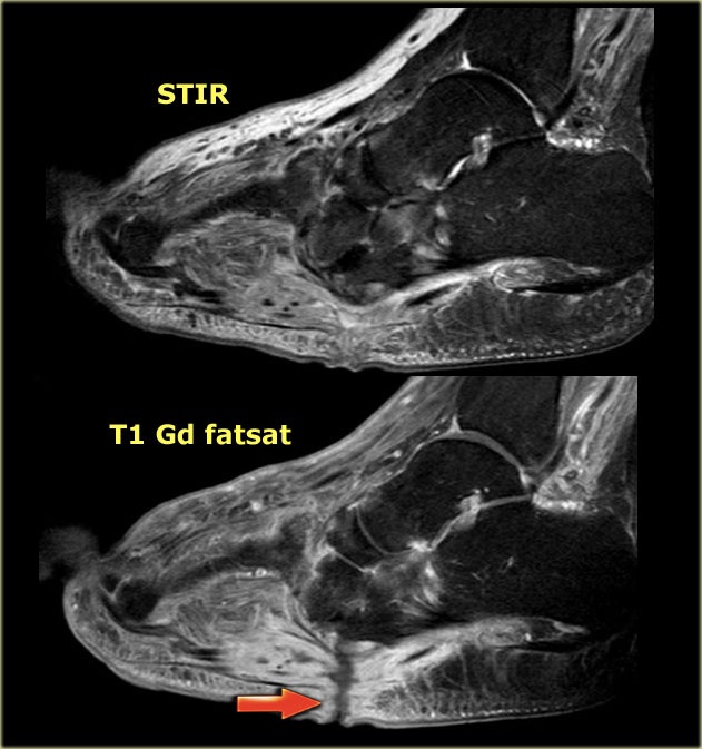
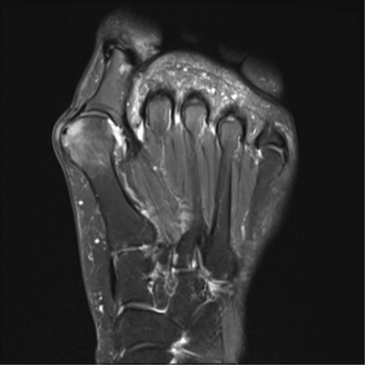
MRI of the left foot. Coronal reformatted T2-WI FS (A) showing subtle …
My Image 92
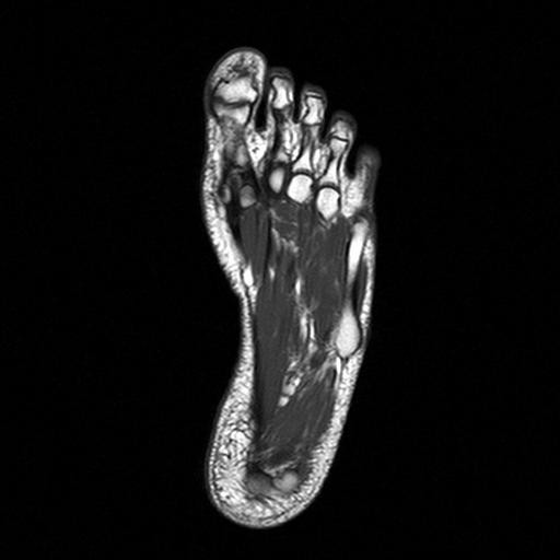
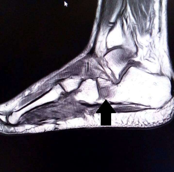
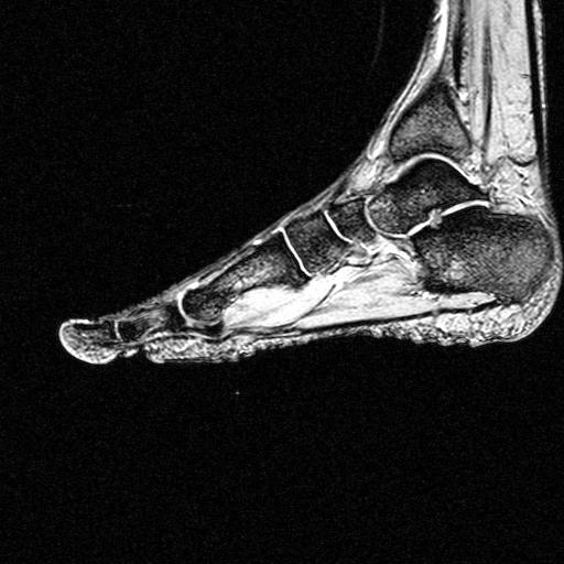
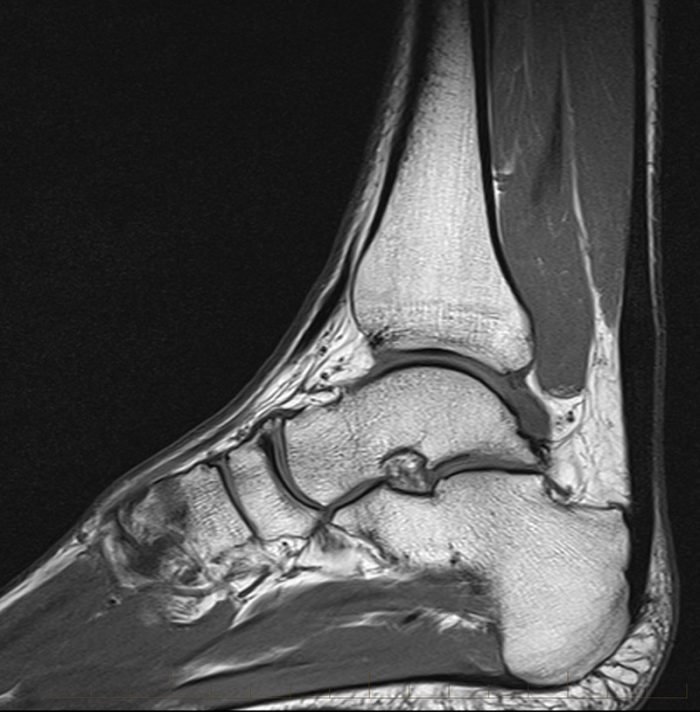
foot mri sagittal t1 image
My Image 93
Feet MRI
My Image 94
Image | Radiopaedia.org
My Image 95
Cases and Images in Infectious Disease
My Image 96
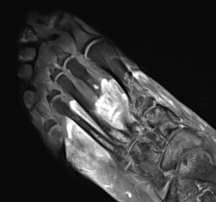
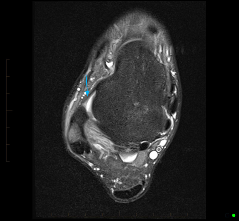
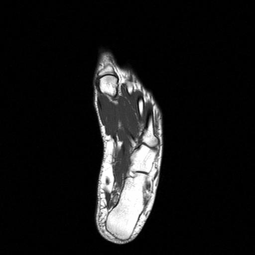
foot mri stir axial image
My Image 97
Pin on MRI
My Image 98
MRI of the foot which showed an image suggestive of bone fractures …
My Image 99
Coloured MRI scan of ankle bones in the human foot – Stock Image – P116 …
We extend our gratitude for your readership of the article about what does an mri of the foot show at finwise.edu.vn. We encourage you to leave your feedback, and there’s a treasure trove of related articles waiting for you below. We hope they will be of interest and provide valuable information for you.