List showcases captivating images of what does a normal mri of the lumbar spine look like finwise.edu.vn
what does a normal mri of the lumbar spine look like
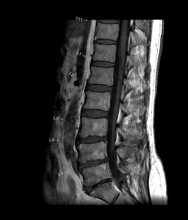
lumbar normal sag mri w text | Mri, Mri technologist, Mri scan
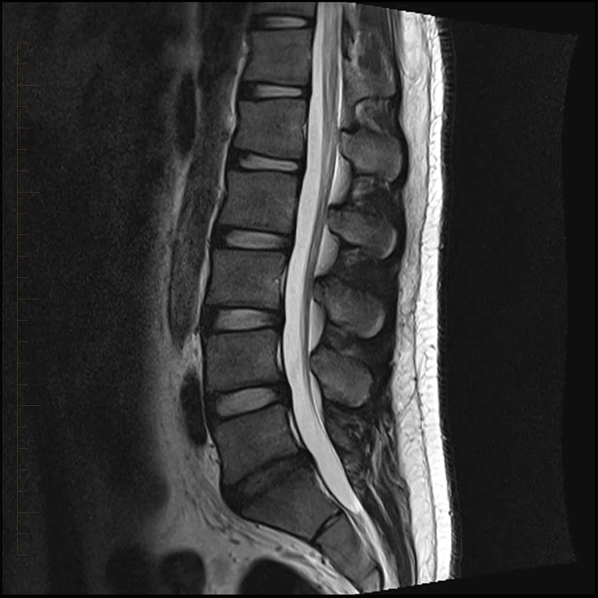
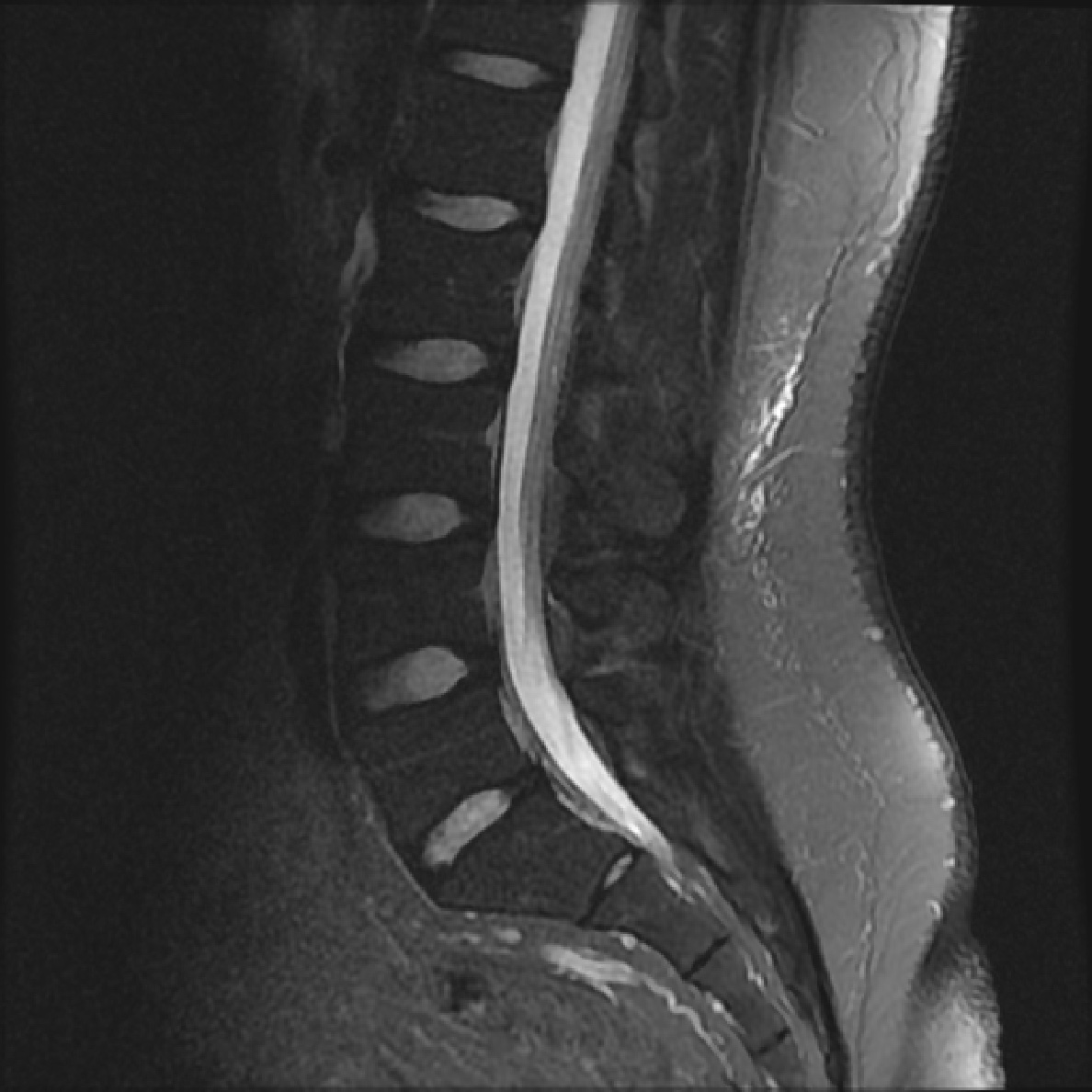
Normal lumbar spine MRI | Image | Radiopaedia.org
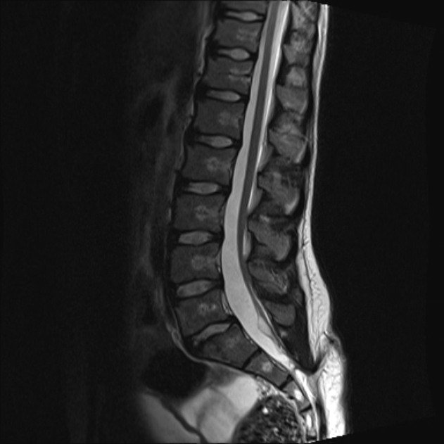
Normal lumbar spine MRI: 3 T | Image | Radiopaedia.org
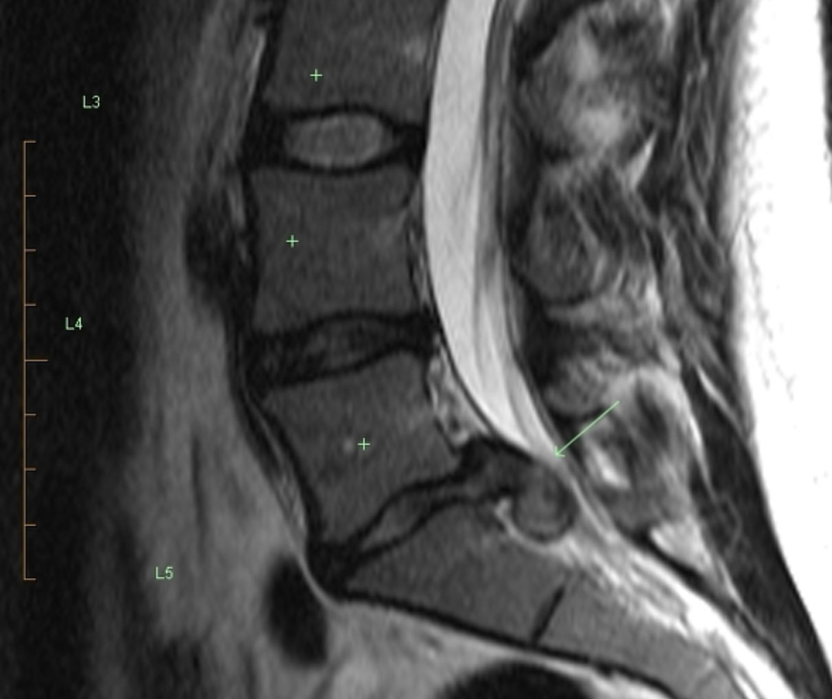
MRI of the Lumbar Spine | Healthy Spine Alignment | Donald Corenman MD …
MRi of lumbar spinal stenosis | Download Scientific Diagram
Full length spine: normal MRI appearance | Image | Radiopaedia.org
Normal lumbar spine MRI: 3 T | Image | Radiopaedia.org
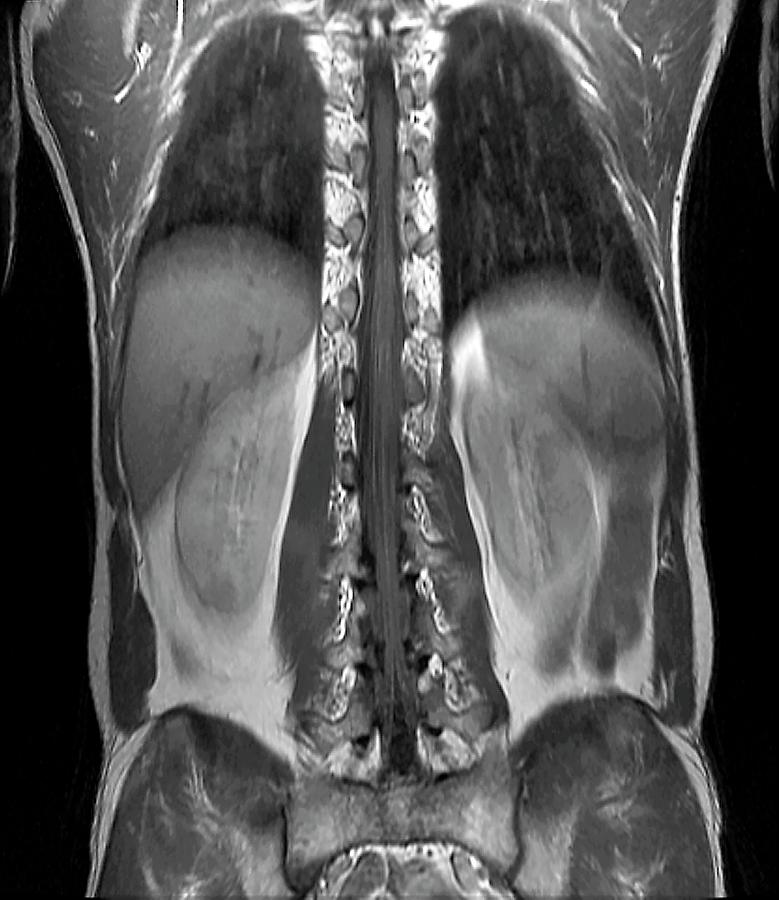
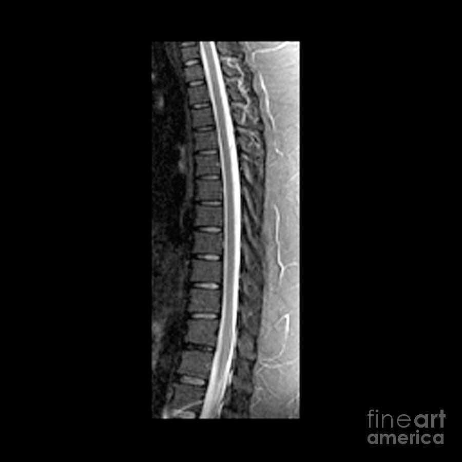
Normal thoracic spine MRI | Image | Radiopaedia.org
b. Normal Sagittal T2 Weighted MR Lumbar Spine | Download Scientific …
Spine Dragon – Spine Basics: Investigations & Imaging
Pin em Spine
MRI of the lumbar spine showed degenerative retrolisthesis of L5 on S1 …
Back Surgery – Clinica SANDALF Benalmadena, Costa del Sol
What does my MRI Mean : What Your MRI Actually Says About You
MRI Scans: Where Abnormal Findings Are Normal – Sohrab Gollogly MD
Pin on X-Ray Lumbar
MRI lumbar spine. (a) MRI of the lumbar spine with contrast showing L2 …
MRI of the Lower Back | Melbourne Radiology
Magnetic resonance imaging (MRI) scan of the lumbar spine eight months …
MRI of the Lower Back – Diagnostic Imaging – Melbourne Radiology
Descriptions of spinal MRI lesions and definition of a positive MRI of …
Normal cervical spine MRI | Image | Radiopaedia.org
Healthcare Extreme How To Read An MRI Lumbar Spine In 8 Easy Steps
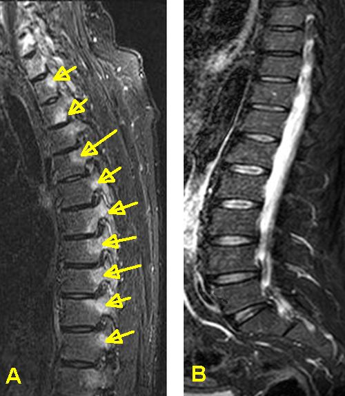
Active Inflammatory Lesions Detected by Magnetic Resonance Imaging in …
Back pain and Normal MRI scan in AS
My lower back MRI results
Healthcare Extreme How To Read An MRI Lumbar Spine In 8 Easy Steps
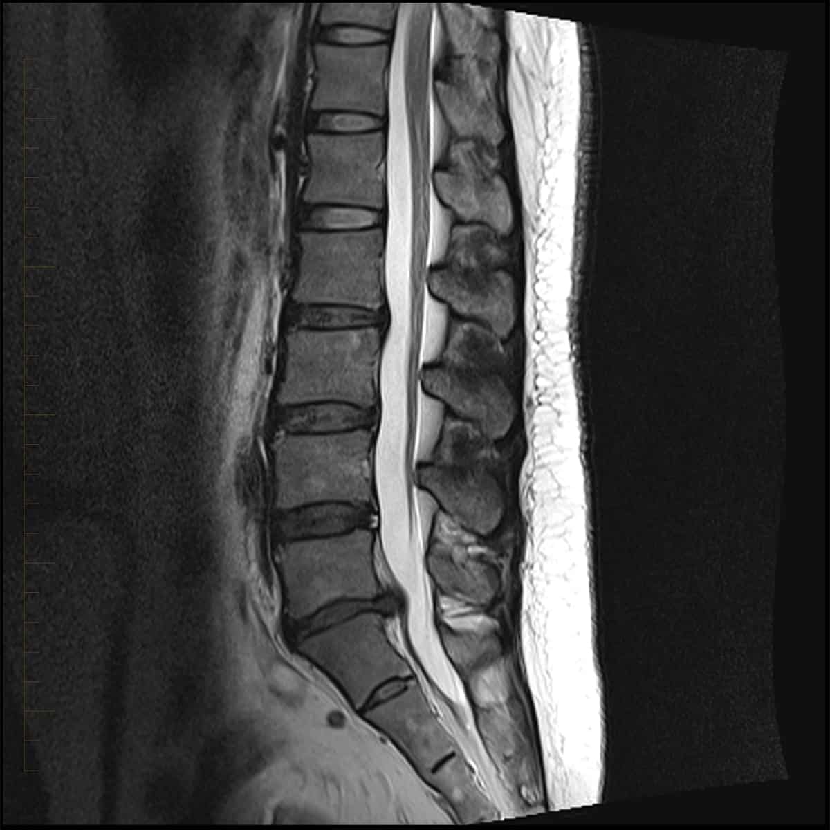
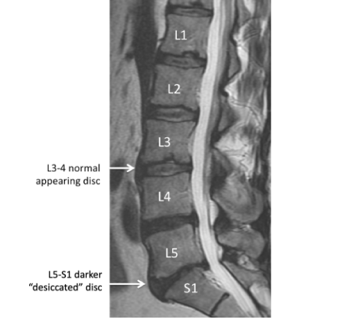
Understanding Your MRI of the Lumbar Spine
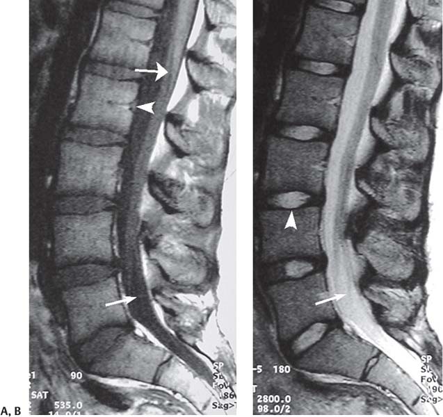
MRI of the lumbar spine. Sagittal (A) and axial (B) T1-weighted MRI of …
Descriptions of spinal MRI lesions and definition of a positive MRI of …
Lumbar spine MRI scan taken four days after posterior lumbar and …
MRI image of human spine | High-Quality Stock Photos ~ Creative Market
Understanding Your MRI of the Lumbar Spine
MRI Lumbar spine 2 view stock illustration. Illustration of …
Preoperative MRI showing moderate spinal stenosis at the L3/4 level (a …
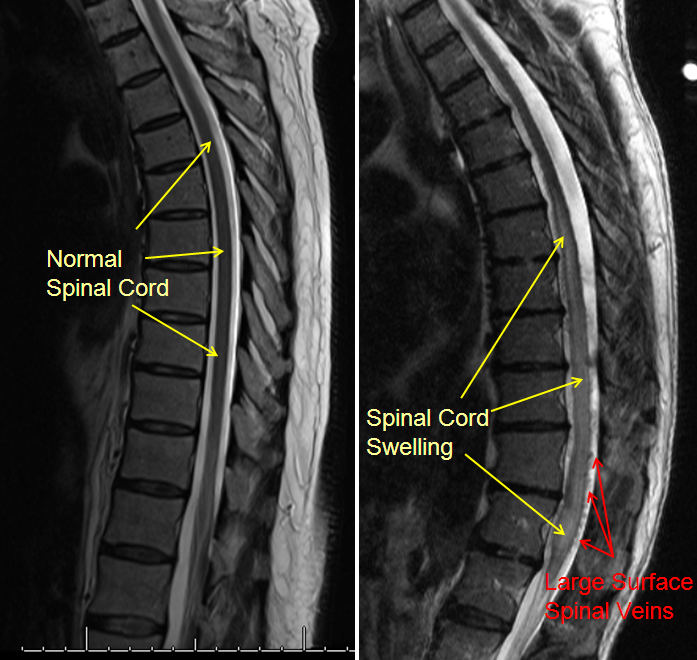
Normal Spinal Cord Photograph by Zephyr
Pin by Kibuuka Brian on KB | Lumbar spinal stenosis, Spinal stenosis …
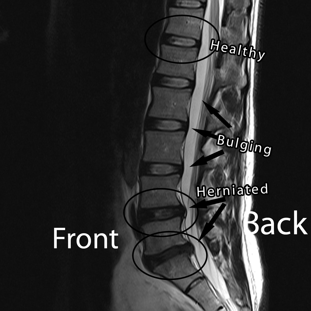
MRI Showing The Lower Back With Healthy Disc Near The Top With The …
MRI of the thoracic spine (T2-weighted, sagittal reconstruction). The …
Magnetic resonance imaging in the prone position and the diagnosis of …
The Radiologist on Instagram: “Sagittal T2 weighted image of an MRI of …
MRI of the Lower Back | Melbourne Radiology
Normal trauma cervical spine MRI | Image | Radiopaedia.org
Normal trauma cervical spine MRI | Radiology Case | Radiopaedia.org
Magnetic Resonance Imaging of the lumbar spine. (A) The T2-weighted …
T2-weighted spinal cord MRI centered on the lumbosacral region: a …
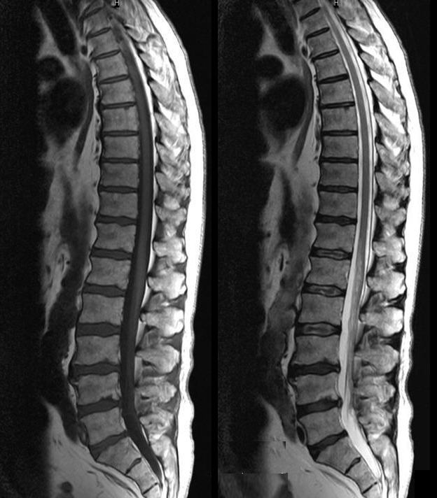
Follow-up MRI lumbar spine, sagittal view. (A) T2 weighted and (B) T1 …
Preoperative sagittal T2-weighted MRI scan of the lumba | Open-i
An MRI of the lumbosacral spine (left) showed an extradural lesion …
The Pediatric Spine | Radiology Key
Healthcare Extreme How To Read An MRI Lumbar Spine In 8 Easy Steps
Figure2.Lumbar spine MRI performed in Case 2. Lumbar spine MRI imaging …
Normal T1 MRI Studies of the Lumbar Vertebral Column
How To Read An Mri Of The Cervical Spine – unugtp
ArUn’s MRI Protocols: MRI Lumbar Spine Protocol
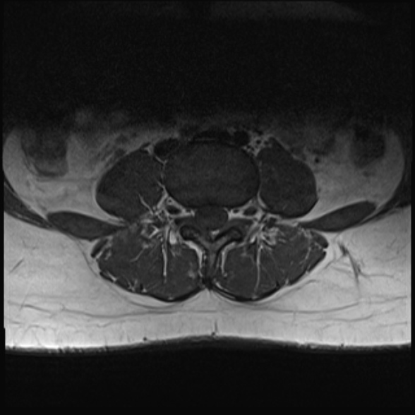
T2-weighted axial MRI scan of the lumbosacral spine showing diminishing …
Healthcare Extreme How To Read An MRI Lumbar Spine In 8 Easy Steps
MRI of a Lumbar-Disk Herniation – License, download or print for £9.92 …
Lumbar Spine: Lumbar Spine Mri With Contrast
Low thoracic and lumbar spine MRI showing a hyperintense lesion in the …
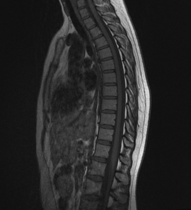
Sagittal Mri Of Normal Thoracic Spine Photograph by Medical Body Scans
Herniated Disc Mri L5 S1 – Lumbar Herniated Disc – A More in Depth Look …
Pin em Back pain
Understanding Your Mri Of The Lumbar Spine | My XXX Hot Girl
Lower Back Injury Prognosis Update | How Do I Get Ripped? | Medical …
Follow-up MRI lumbar spine, sagittal view. (A) T2 weighted and (B) T1 …
Sagittal T2 weighted MR image of the thoracic and lumbar spine …
Normal trauma cervical spine MRI | Radiology Case | Radiopaedia.org
MRI of Lumbar spine | Magnus MRI & Ultrasound Diagnostic Center 磁聲磁共振及 …
Image Gallery normal cervical mri
Image | Radiopaedia.org
MRI Lumbar Spine | GWIC
MRI scans showed normal spinal cord and intervertebral discs. MRI also …
MRI Spine – Paediatic MRI Series
MRI lumbosacral spine of the patient showing mild to moderate lumbar …
slipped disc lumbar x ray – Pippa Dowd
Follow up post contrast cervical spine MRI. Parasagittal and axial T1w …
A T2-weighted sagittal view of the whole spine MRI of t | Open-i
Lumbar Spine: Ct Myelogram Lumbar Spine
Talking to Patients About Pain Can Improve Lumbar Spine MRI …
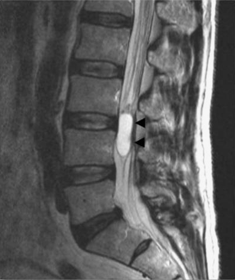
Cureus | Isolated Lumbar Spinal Nerve Root Myxopapillary Ependymoma
7 Problems with Imaging – Open Health Clinic
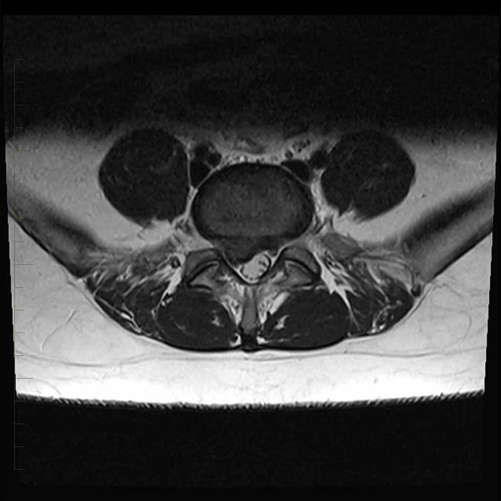
Lumbar MRI, axial T2 sequence without contrast enhancement, showing …
Spinal Dural Fistula | neuroangio.org
Lumbar Spine MRI: (A) sagittal T2WI showing degenerative disc changes …
check out my Xrays and tell me what you think – General Discussion …
Dr Balaji Anvekar FRCR: Multiple Myeloma MRI Spine
9 Lumbar spine MRI showing tethered spinal cord with conus medullaris …
Normal Spine Mri | www.pixshark.com – Images Galleries With A Bite!
1 Lumbar spine imaging of a 72-year-old female patient with lower back …
VIDEO
MRI or USG for pain | दर्द में कौनसी जाँच सबसे बेहतर ? | Dr Rachit Gulati | SAAOL Ortho Care