List showcases captivating images of ovarian cancer pictures on ultrasound finwise.edu.vn
ovarian cancer pictures on ultrasound
Ovarian Clear Cell Adenocarcinoma | Узи
Ovarian Cancer Ultrasound Of Ovaries With Cyst : Sonographic Assessment …
Ovarian Granulosa Cell Tumor – OB/GYN Case Studies – CTisus CT Scanning
Imaging in gynecological disease (12): clinical and ultrasound features …
Ovarian Serous Adenocarcinoma (mainly necrotic) | Medical ultrasound …
Imaging in gynecological disease (11): clinical and ultrasound features …
New sonographic marker of borderline ovarian tumor: microcystic pattern …
Ovarian Granulosa Cell Tumour | Medical ultrasound, Obstetric …
Screening for ovarian cancer | The Medical Journal of Australia
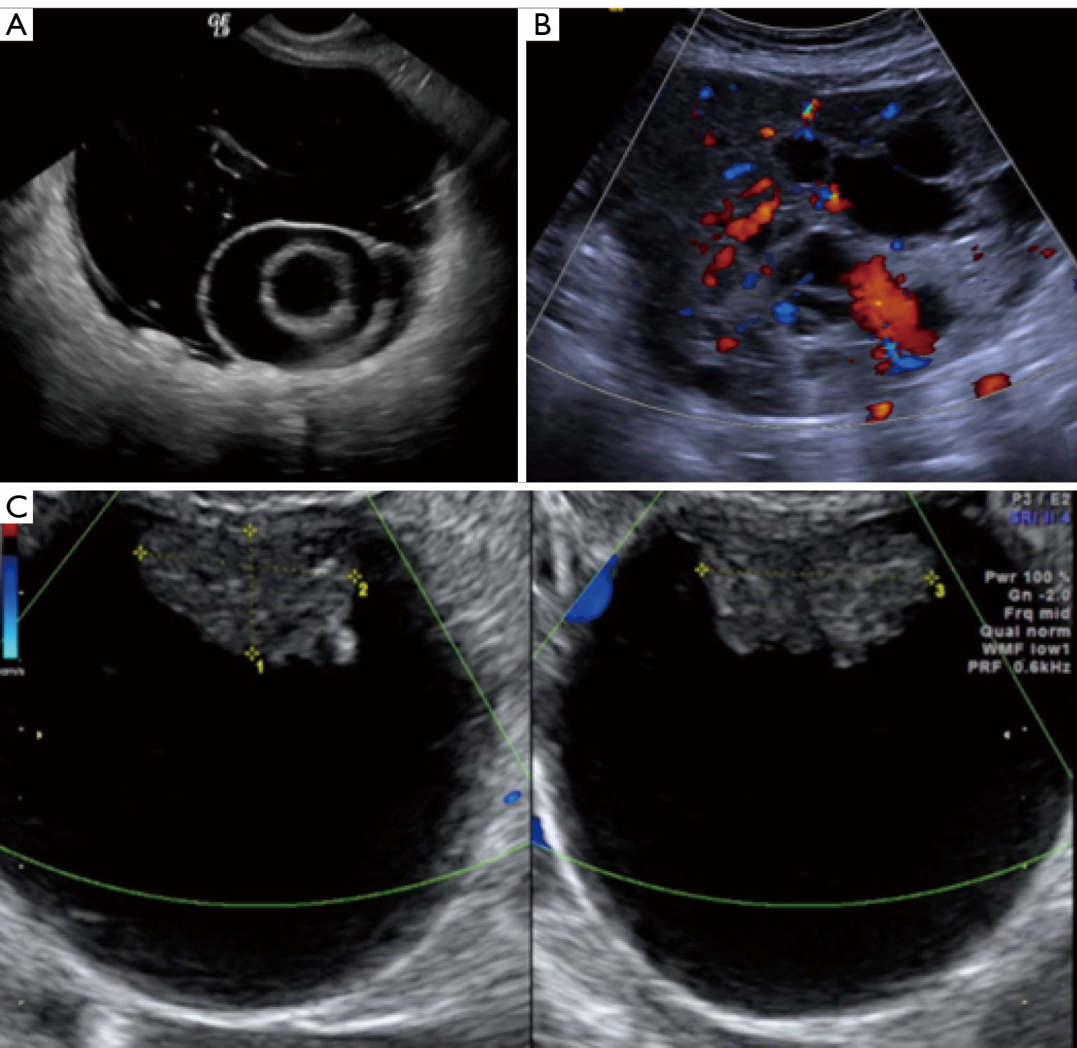
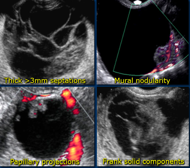
Ovarian masses with papillary projections diagnosed and removed during …
Ovarian Serous Adenocarcinoma | Educational websites, Gynecology …
Ovarian masses with papillary projections diagnosed and removed during …
Preoperative sonographic features of borderline ovarian tumors …
Cyst On Ovaries Ultrasound | My XXX Hot Girl
See all 34 photos
What Does Ovarian Cancer Look Like On An Ultrasound – What Does
Ovarian cancer, MRI scan – Stock Image – C050/9705 – Science Photo Library
VIETNAMESE MEDIC ULTRASOUND: CASE 302 : OVARY TUMOR , Dr PHAN THANH HẢI …
Ovarian Malignant Germ Cell Tumors: Cellular Classification and …
Ovarian Tumors (Clinical Setting and US) | Radiology Key
A simple ovarian cyst on the right side of the uterus fulfilling all …
Ovarian Tumors (Clinical Setting and US) | Radiology Key
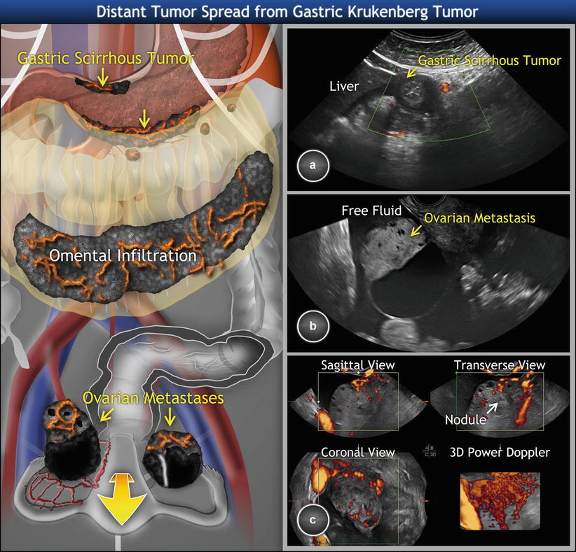
Recurrent ovarian cancer in a 49year-old female patient with ovarian …
Ovarian Cancer Ultrasound Vs Normal : The Characteristic Ultrasound …
Early Diagnosis of Ovarian Carcinoma: Is a Solution in Sight? | Radiology
Ovarian Cancer Ultrasound Of Ovaries With Cyst : Sonographic Assessment …
Ovarian Malignant Germ Cell Tumors: Cellular Classification and …
Ultrasound image of ovary with follicles | Download Scientific Diagram
Ovarian Cancer with Carcinomatosis – OB/GYN Case Studies – CTisus CT …
MRI scan of early-stage 1A high-grade serous ovarian cancer. The …
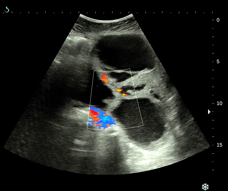
Transabdominal ultrasound showing a signifi cant amount of ascites in a …
Ovarian Cancer Ultrasound Vs Normal : The Characteristic Ultrasound …
Ultrasound characteristics of early-stage high-grade serous ovarian …
Ovarian Cancer Ultrasound Of Ovaries With Cyst : Sonographic Assessment …
Ovarian cysts, ultrasound scan – Stock Image – M850/0652 – Science …
(PDF) Ultrasound and Clinical Preoperative Characteristics for …
Pin on adnexa adnexa
Ovarian torsion with dermoid cyst | Image | Radiopaedia.org
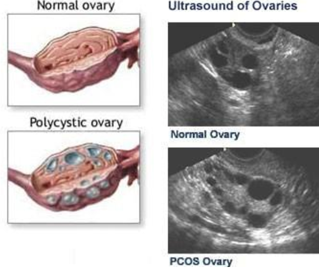
What’s Polycystic Ovarian Syndrome? | Scienceline
Selection of hens with normal ovaries or ovarian tumors using (A-C …
📃 Ovarian cyst
Polycystic Ovarian Syndrome (PCOS) – Davies Chicago Fertility …
Ovarian Teratomas: Tumor Types and Imaging Characteristics | RadioGraphics
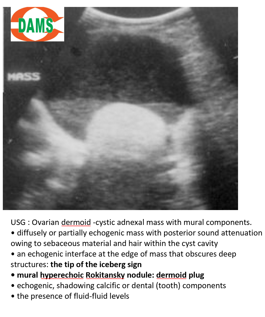
Ovarian dermoid cyst with Rokitansky nodule | Radiology Case …
Extremely large epithelial ovarian cancer associated with pregnancy: A …
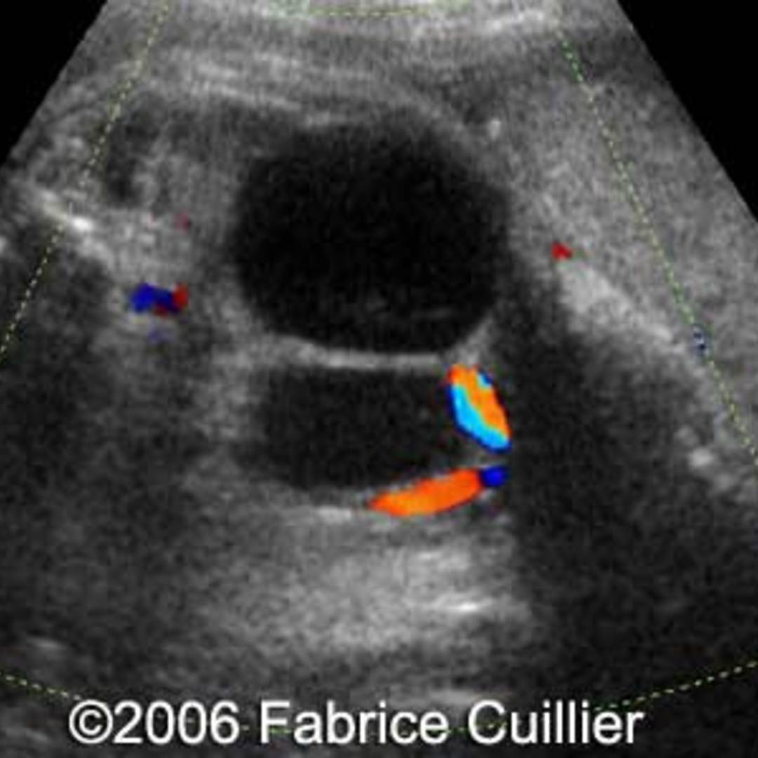
Ultrasonographic color Doppler appearance of the ovaries with moderate …
Simple ultrasound‐based rules for the diagnosis of ovarian cancer …
Ovarian cancer, MRI scan – Stock Image – C050/9706 – Science Photo Library
Place of Doppler Ultrasound in the Characterization of Ovarian Cysts …
(PDF) Enhancement of Ovarian Tumor Detection With αvβ3 Integrin …
Bilateral ovarian cancer, MRI scan – Stock Image – C034/5656 – Science …
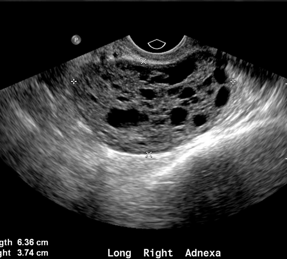
The characteristic ultrasound features of specific types of ovarian …
Ovarian Cysts – Womens Ultrasound ClinicWomens Ultrasound Clinic
Ovarian Malignant Germ Cell Tumors: Cellular Classification and …
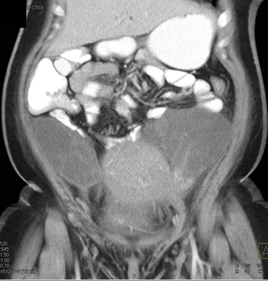
CT scan showing adnexal mass adjacent to left ovary. | Download …
mage of the ovarian tumor on transvaginal ultrasound examination …
Polycystic Ovarian Syndrome: Role of Imaging in Diagnosis | RadioGraphics
Ultrasound Evaluation of Ovaries | Obgyn Key
Ovarian Granulosa Cell Tumor – OB/GYN Case Studies – CTisus CT Scanning
Ovarian Tumors (Clinical Setting and US) | Radiology Key
Left: Polycystic ovarian morphology using HDlive without (upper left …
Ovarian Tumors (Clinical Setting and US) | Radiology Key
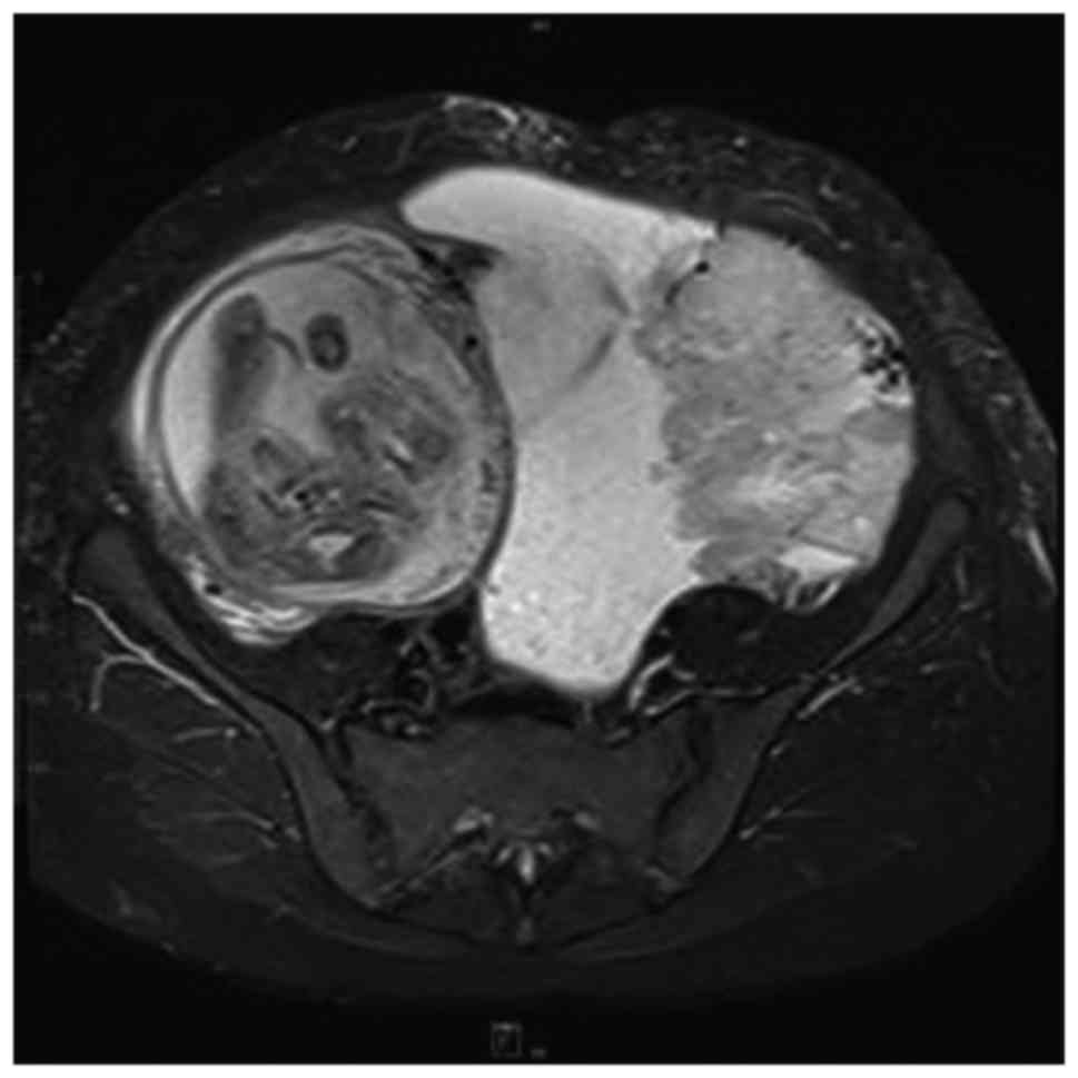
MRI in ovarian cancer
Ovarian ectopic pregnancy. Transvaginal ultrasound showing a …
Ovarian Dermoid: Ultrasound Teaching File – Sumer’s Radiology Blog
Ovarian Granulosa Cell Tumor – OB/GYN Case Studies – CTisus CT Scanning
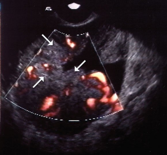
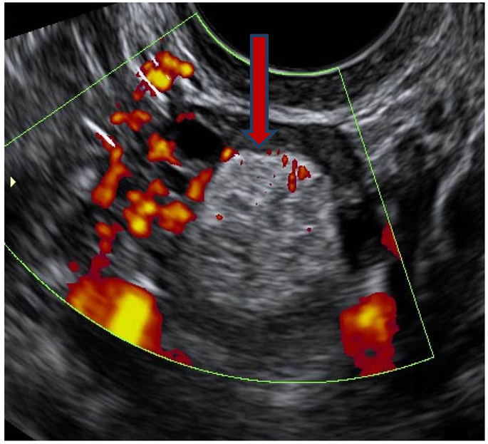
a Measurement of ovarian tumor by 2DUS. b 2DUS with color Doppler …
Rt ovarian torsion in 13 y old f | Ultrasound, Sonography, Radiology
Sonographic images of benign and malignant ovarian morphology. Numeric …
What To Do If You’ve Been Diagnosed With PCOS | HuffPost
Ovarian Cysts – Dr. Elena Rodriguez
Ovarian Carcinoma with Bilateral Adnexal Masses – OB/GYN Case Studies …
Transvaginal ultrasound of the ovary. Note ovarian stroma and …
Ovarian cancer, MRI scan – Stock Image – C050/9703 – Science Photo Library
Ultrasound image gallery | Ovaries, Image, Ovarian
VIETNAMESE MEDIC ULTRASOUND: CASE 526: BIG OVARIAN CYSTIC TUMOR, Dr …
Pin on Ultrasound
Management of ovarian cysts | UW Ultrasound
granulosa cell tumor in kids on ultrasound – would cause preocious …
Types of cervical cancer Archives – Ultrasoundfeminsider
Non-visualization of the ovary on CT or ultrasound in the ED setting …
Ovarian Cancer | CancerQuest
Pin on Radiology
Pin on ovarian cancer awareness
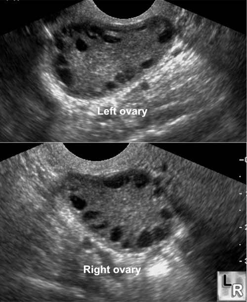
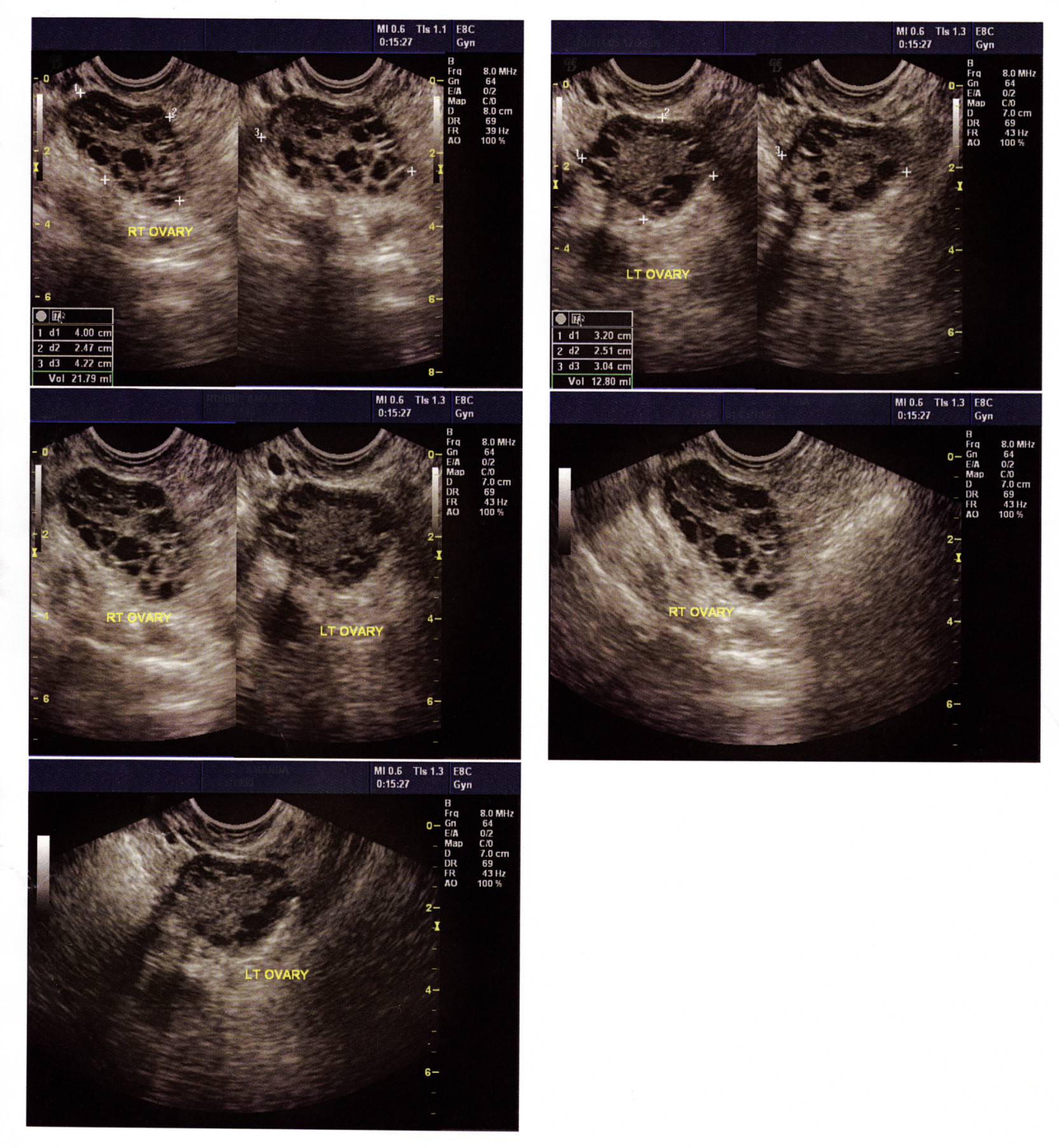
Ultrasound images showing enlarged right and left ovaries with multiple …
Transvaginal ultrasound images showing typical features of a right …
Ovarian Torsion | Diagnostic medical sonography, Ultrasound sonography …
Functional ovarian cysts | Download Scientific Diagram
Pin on ovarian tumors
Ovarian Granulosa Cell Tumor – OB/GYN Case Studies – CTisus CT Scanning
Film Ultrasound Ovarian Cystsinternal Organs Examination For Women …
Film Ultrasound, Ovarian Cysts,Internal Organs Examination For Women …
Ovarian mass in ultrasound. | Download Scientific Diagram
Small cell lung cancer metastatic to the ovary diagnosed during …
Accuracy of ultrasound subjective ‘pattern recognition’ for the …
Borderline ovarian serous tumor (HE staining, ×100). | Download …
Ovarian Endometrioma (ULTRASOUND PELVIS) | Download Scientific Diagram
Cureus | A Comparison of the Clinical Presentation of Ovarian …
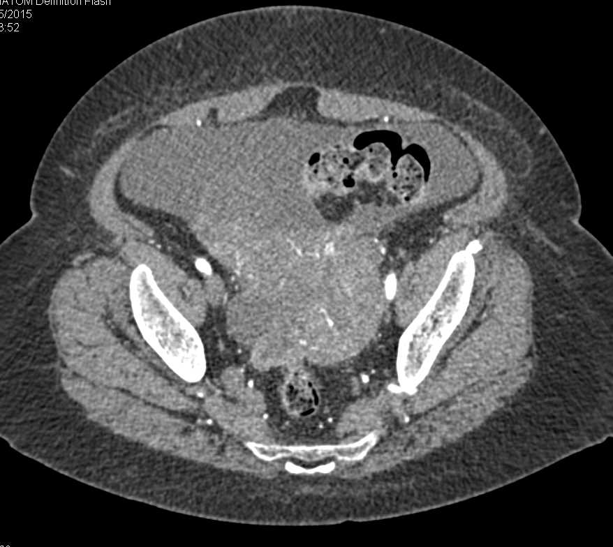
What Does Ovarian Cancer Look Like On A Ct Scan – What Does
(PDF) Ultrasound manifestations of lobulated ovaries: Case report
Figure 2 from MRI features of primary and metastatic mucinous ovarian …
Ovarian masses with papillary projections diagnosed and removed during …
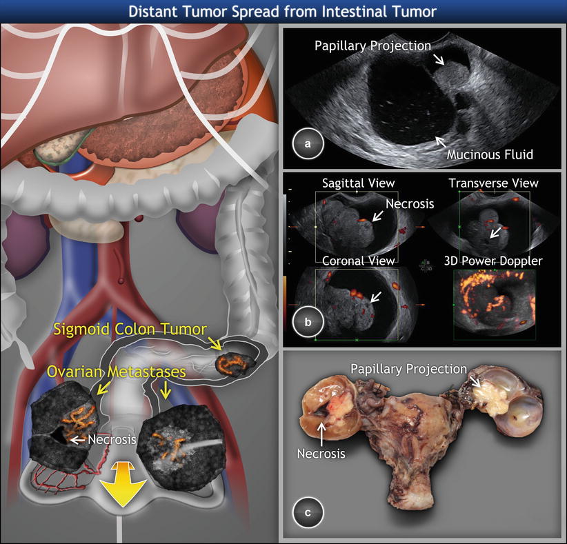
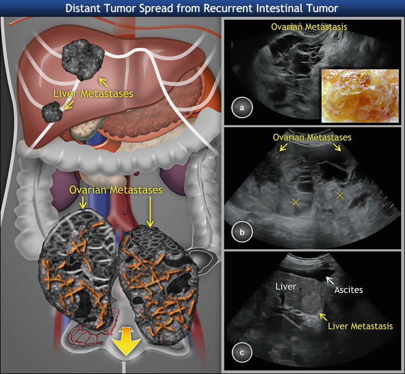
Recurrent intestinal carcinoma involving the ovaries. (a) Transvaginal …
We extend our gratitude for your readership of the article about
ovarian cancer pictures on ultrasound at
finwise.edu.vn . We encourage you to leave your feedback, and there’s a treasure trove of related articles waiting for you below. We hope they will be of interest and provide valuable information for you.