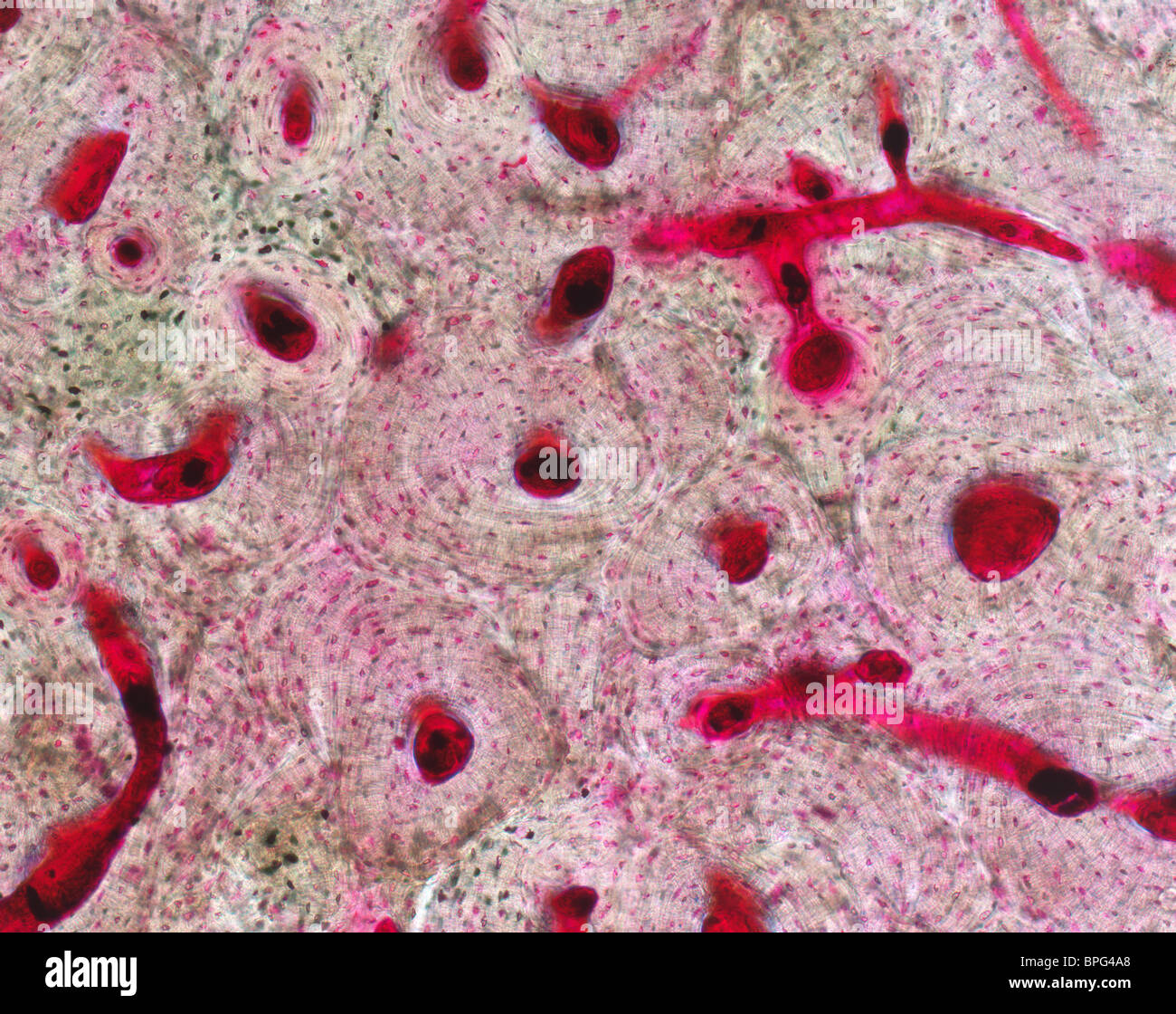
Collection showcases captivating images of concentric lamellae within an osteon are connected by lacunae. finwise.edu.vn
concentric lamellae within an osteon are connected by lacunae.
structure of compact bone Diagram | Quizlet
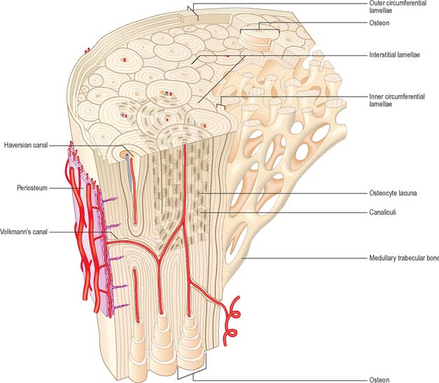
This photo shows a model of an osteon. It points out the blood vessels …
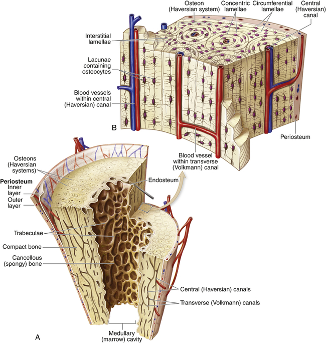
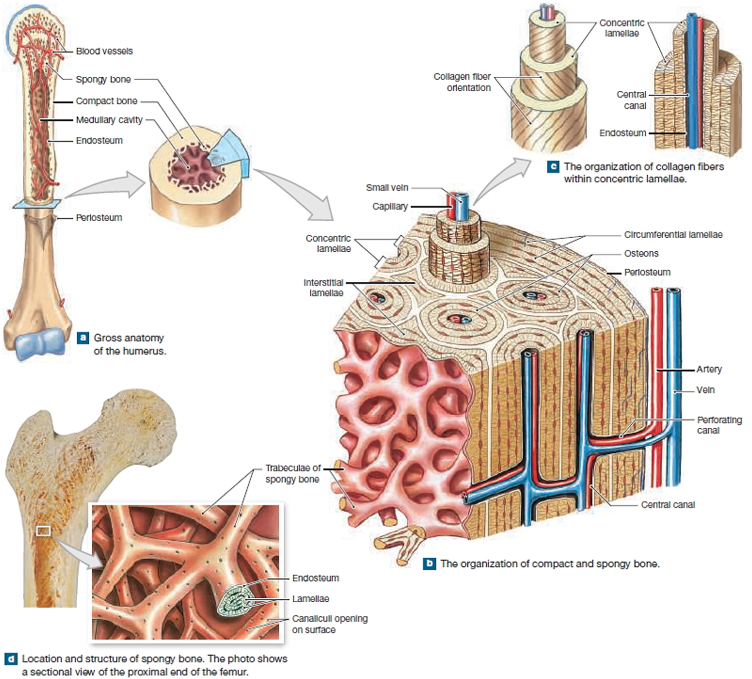
2: Compact bone organisation Top = The organisation of osteons and …
Illustration of compact bone showing the relationship between …
is the inner and outer circumferential lamellae is concentric or no
Regions and Structures of an Osteon Diagram | Quizlet
2 Cross-section of bone showing a cortical bone and spongy/cancellous …
Label The Photomicrograph Of Compact Bone. – Label The Photomicrograph …
Anatomy and Physiology 1 Chapter 6 Bone Tissue Flashcards | Quizlet
Osteon Diagram
Microscopic structure of Compact and Spongy bone tissue Diagram | Quizlet
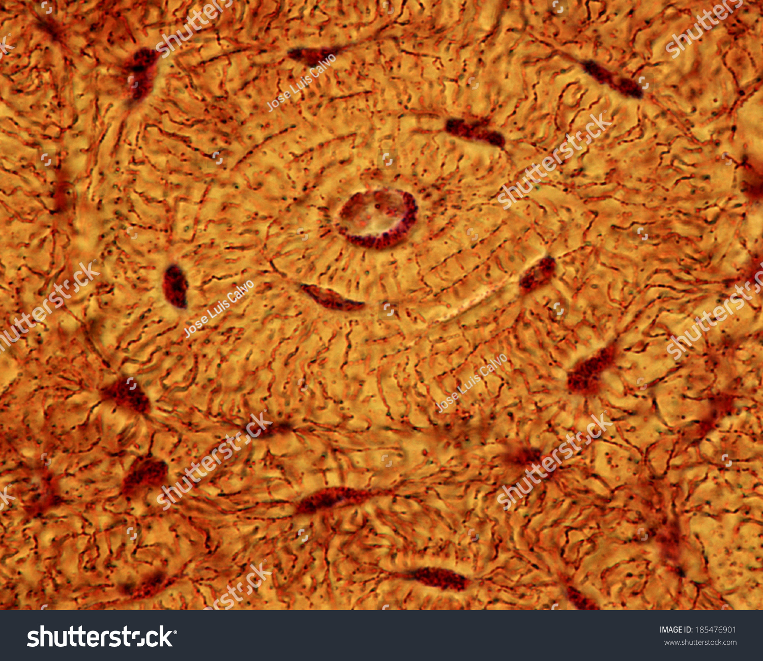
Microscopic structure of human lamellar bone. 35 | Download Scientific …
Histology Of Compact Bone Diagram / Module 6.2 Microscopic Structure of …
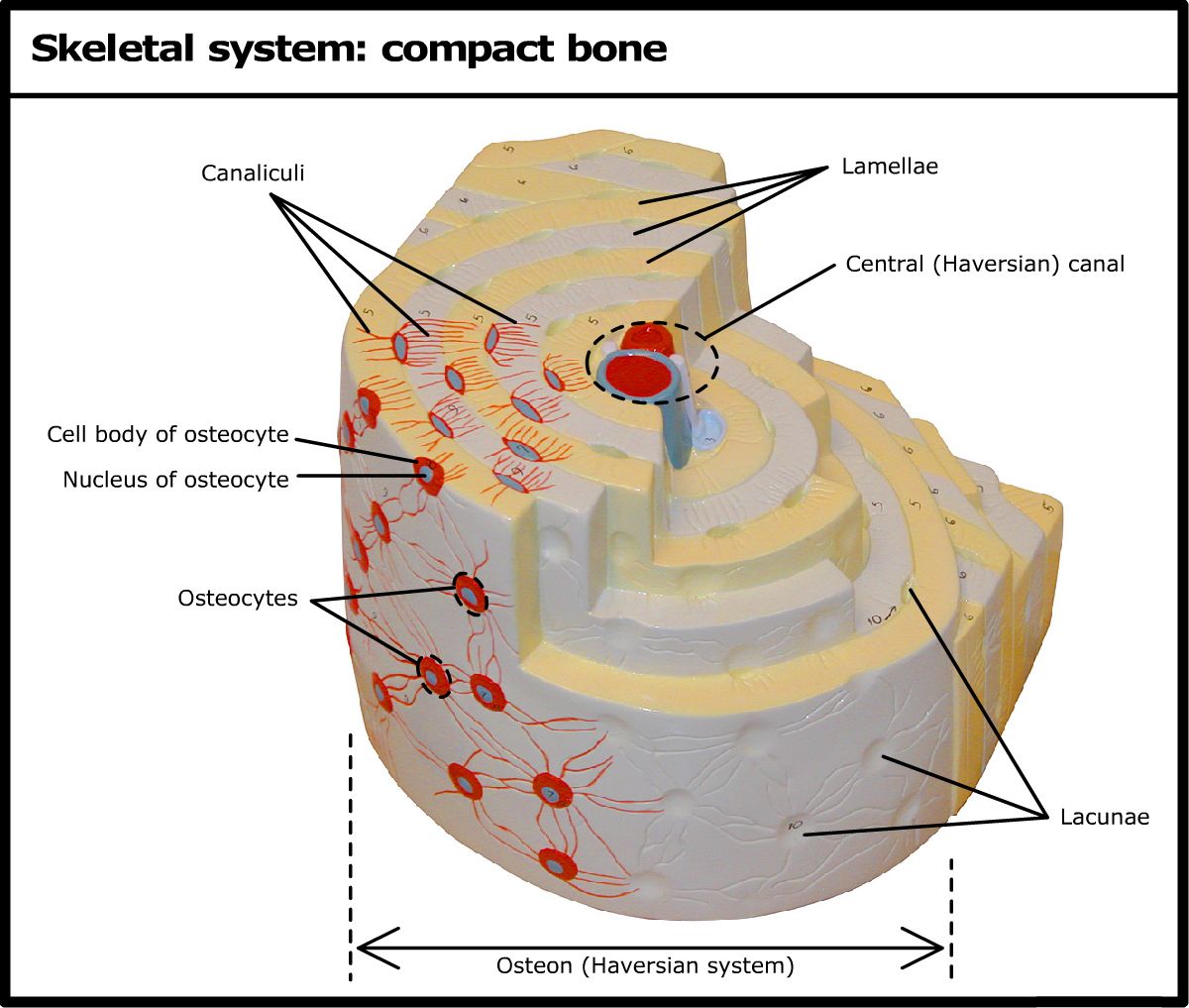
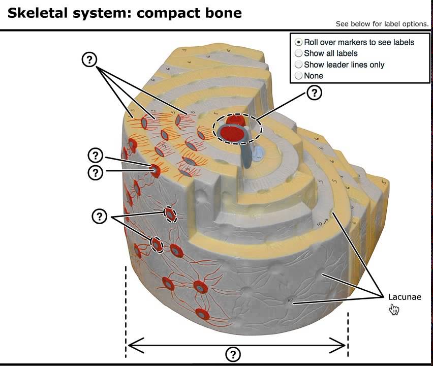
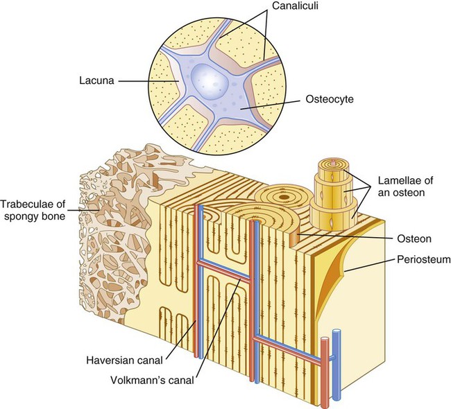
Skeletal system: compact bone
Lacuna Anatomy
(A) Histological cross-section of cortical bone, showing osteon with …
Spongy And Compact Bone Diagram / Diagram Human Bone Anatomy Useful …
Pin on Anatomy and Physiology
33.2C: Connective Tissues: Bone, Adipose, and Blood – Biology LibreTexts
Osteon of Human Bone – YouTube
Osseous Stracture Human Body : Human Body | General Science Quiz …
30 In The Diagram, Where Is The Osteon_ – Wiring Diagram Database
Osteon structure scheme. a = Haversian canal diameter; b = osteon …
Lamella, Lacuna, Canaliculus, Central Canal, Matrix, Bloo…
6: Bone | Pocket Dentistry
Bones at Liberty High School – StudyBlue | Anatomy and physiology …
A. Secondary Osteon and B. Microscopic Structure of Mature Long Bone …
Lacuna Definition and Examples – Biology Online Dictionary
Bones: Structure and Types | Concise Medical Knowledge
1: a) Osteon, formed by bone lamellae with the haversian channel in the …
Lamellae Definition Anatomy – Anatomical Charts & Posters
Compact Bone Diagram Lacunae : Bone Histology General Overview Compact …
Secondary Osteon
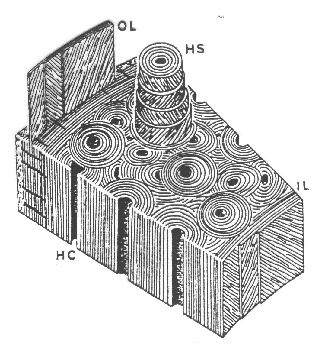
6 A single osteon. The osteon is drawn as if pulled out like a …
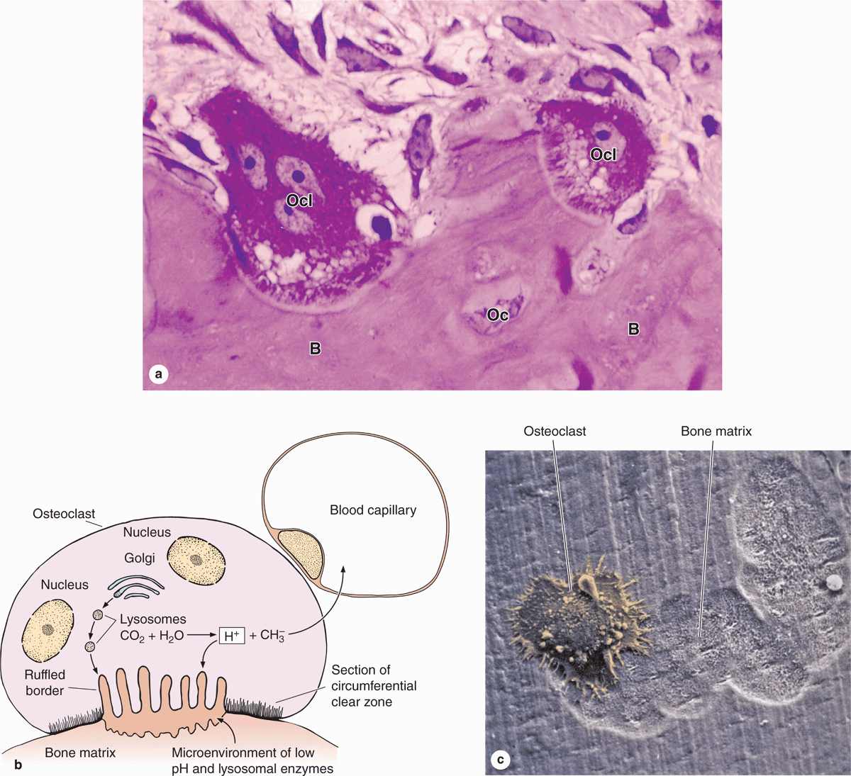
Osteoblasts, Osteoclasts, Calcium, and Bone Remodeling | Owlcation
Osteon Bone Haversian Blood Vessels | Art In Anatomy
Compact Bone Diagram Lacunae – Cartilage Bone Ossification The …
Which Of The Following Labels Best Matches Osteocyte 15+ Pages Solution …
Osteon haversian system, lamellae, blood vessel continues…
Osteon Anatomy
Osteon Bone Haversian Lacunae Canaliculi | Art In Anatomy
(A) Histological cross-section of cortical bone, showing osteon with …
Study Notes
Anatomy lab exam Tissues Flashcards | Easy Notecards
Functional anatomy of the musculoskeletal system | Basicmedical Key
Bone | Basicmedical Key
Structure Of Compact Bone And The Osteon – sharedoc
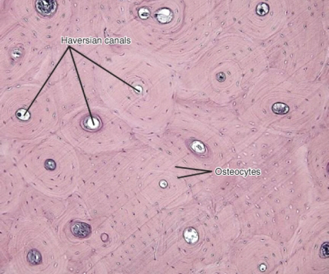
Light Micrograph Of An Osteon In Compact Bone Showing Osteocyte Lacunae …
Solved T u engell lamelle Concentric of Interst n lamellal | Chegg.com
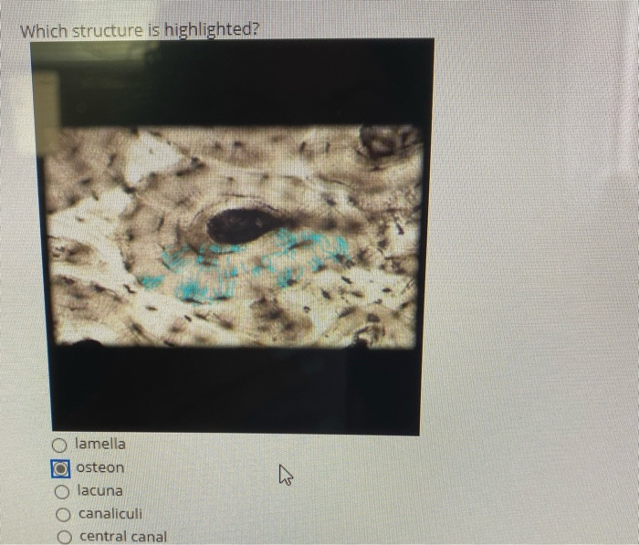
Solved Which structure is highlighted? 2 lamella osteon | Chegg.com
osteon – Liberal Dictionary
Micrografía de una luz de hueso mostrando las laminillas concéntricas …
Histology Glossary: Histology – Compact Bone | Draw It to Know It
Cartilage/Bone/Muscle Histology Notes – Medical Histology – Jacobs …
Micrografía de una luz de hueso mostrando las laminillas concéntricas …
Activity 4: Examining the Microscopic Structure of Compact Bone …
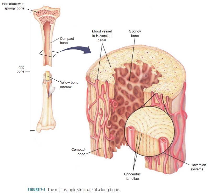
6.4: Compact bone contains parallel osteons, and spongy bone contains …
Compact Bone Diagram Class 9 : Diffusion/Osmosis at University of …
Are there holes in bones in order to let blood cells out? If not, how …
A photomicrograph of an osteocyte embedded in its lacuna. Evolved from …
Structure and Function of the Musculoskeletal System | Basicmedical Key
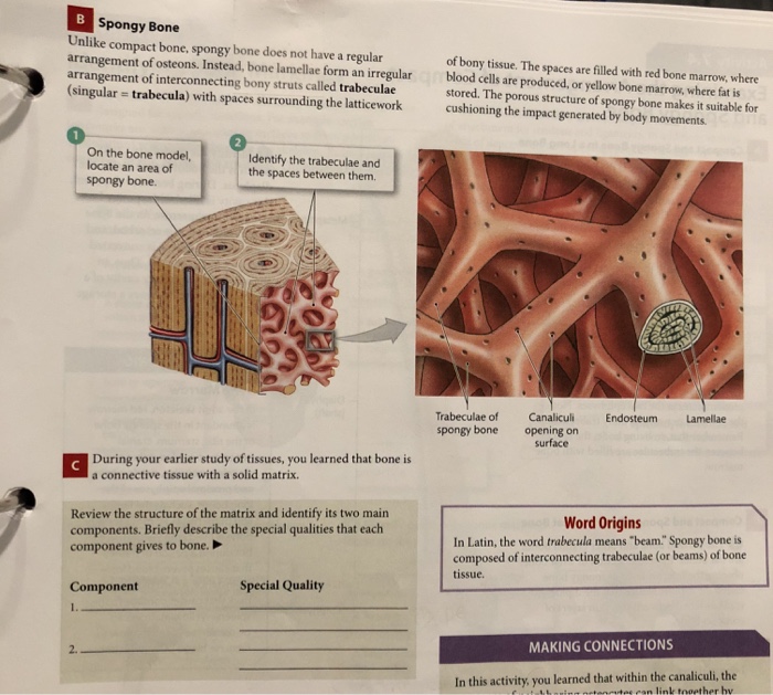
Solved Spongy Bone Unlike compact bone, spongy bone does not | Chegg.com
Hormonal Control of Calcium & Phosphate Metabolism & the Physiology of …
Overview of the osteocyte network within an osteon. a) 3D rendering of …
Structure and Function of the Musculoskeletal System | Basicmedical Key
Bone Bruise – Causes, Symptoms, Diagnosis, Treatment & Healing Time
Microscopic Anatomy (Bone Cells) – Structures of Bones
[Solved] Label the following on the Ground compact bone slide 1 …
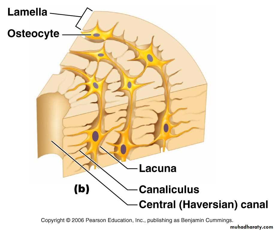
bone tissue pptx – dr.suhaila – Muhadharaty
10 Concentric and interstitial lamellar bone (transverse section-ovine …
MCQs on Cell, Tissue, Skeleton System and Joint Vipin Vageria
Anatomy 312 > Brad Seebach > Flashcards > Exercise 6 – Histology: Bone …
The Skeletal System ‹ OpenCurriculum
Print Chapter 6: Osseous Tissue and Bone Structure flashcards | Easy …
Solved Label the photomicrograph of compact bone. Osteocyte | Chegg.com
6: Describe the structure of the osteon in cortical bone AND 7: Explain …
A-C Ultrastructure of bone. A Light microscope image of a ground …
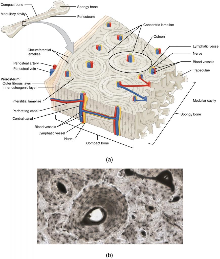
6.3 Bone Structure — Anatomy and Physiology – Anatomy and Physiology …
Histology – Spongy Bone | Nursing notes, Osteology, Bones
Compact Bone Diagram Pearson : Medullary Cavity Wikipedia / What is the …
In The Diagram Where Is The Osteon – Wiring Diagram
11 Acoustic impedance images of human cortical bone cross-sections from …
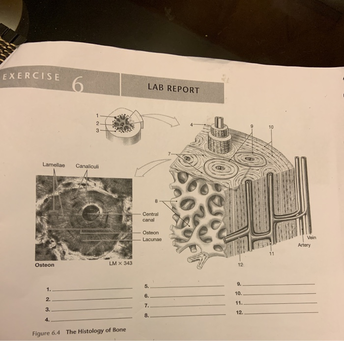
Solved EXERCISE 6 LAB REPORT Lamellae Canaliculi Central | Chegg.com
The lamellar units consist of concentric layers of elastic lamellae …
Study Notes
Schematic of osteon. A . Cross section. Indicated are Haversian canal …
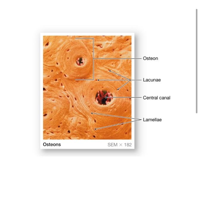
Solved Osteon Lacunae Central canal Lamellae Osteons SEM X | Chegg.com
(a) Basic bone biology. In (a), (A) macroscopically, bone appears as …
Model of fibrillar orientation relative to the osteon axis. The …
Compact Bone Diagram Lacunae / Structure of compact bone. | Download …
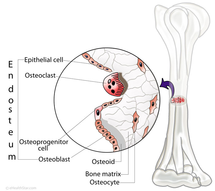
Endosteum Definition, Function, Location, Structure, Pictures | eHealthStar
Study Notes
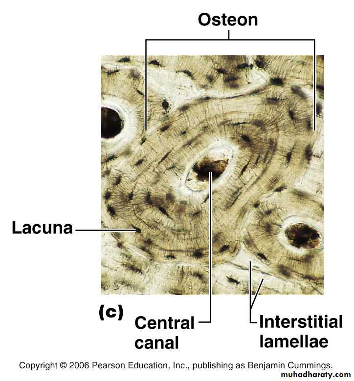
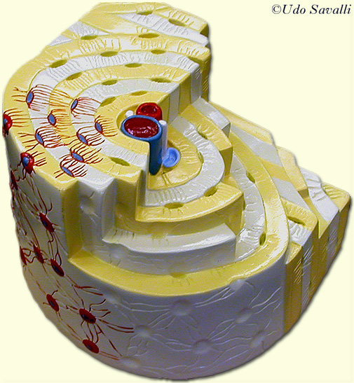
bone tissue pptx – dr.suhaila – Muhadharaty
Cross Section Of A Compact Bone – File Transverse Section Of Bone Svg …
BIO201-Bone
2 Lamellae in a circularly polarized microscopy image of human cortical …
3D Skeletal System: Compact Bone, Spongy Bone, and Osteons—Oh My!
Schematic of cell wall layers within cellular structure of wood. The S2 …
Examples of woven tissue and primary osteons within diaphyseal …
Bone Structure
Pin su Chapter 7 – Bone Tissue